


Page 3
Page 3

Functional imaging and modeling of the heart ; 3rd International workshop, FIMH 2005, Barcelona, Spain, June 2-4, 2005, Proceedings
The FIMH conference was the ?rst attempt to agglutinate researchers from several complementary but often i- lated ?elds: cardiac imaging, signal and image processing, applied mathematics and physics, biomedical engineering and computer science, cardiology, radi- ogy, biology, and physiology. In the ?rst two editions, the conference received an enthusiastic acceptance by experts of all these communities. FIMH was ori- nally started as a European event and has increasingly attracted more and more people from the US and Asia. This edition of FIMH received the largest number of submissions so far with a result of 47 papers being accepted as either oral presentations or posters. There were a number of submissions from non-EU institutions which con?rms the growing interest in this series of meetings. All papers were reviewed by up to four reviewers. The accepted contributions were organized into 8 oral sessions and 3 poster sessions complemented by a number of invited talks. This year we tried to allocate as many papers as possible as oral presentations to facilitate more active participation and to stimulate multidisciplinary discussions.

Fractal Behaviour of the Earth System
In this volume a collection of - pers considers the fractal behavior of the Earth's continental crust. Surface gravity anomalies are known to exhibit power-law spectral behavior under a wide range of conditions and scales. La Manna utilize multifractal models to explain the behavior of well logs from the main KTB borehole in Germany.

Essentials of Restenosis : For the Interventional Cardiologist
Essentials of Restenosis provides the practicing cardiologists and biologists an elaborate review of the current advances in the field. The book explore the process of restenosis from bench to bedside, encompassing in equal detail the newest scientific findings of the biological mechanisms as well as the progression in development of diagnostics and treatments. The volume also provides the reader with a basic understanding of the biological process and the latest diagnostics and treatments of the disease, with an up-to-date detailed discussion of the most important recent findings.

Emergency Neuroradiology
The resulting work is an up-to-date technical tool that preserves its original aim of contributing to the training of those radiologists who work in emergency departments.

Electronic Devices and Circuit Design : Challenges and Applications in the Internet of Things
Offers a broad view of the challenges of electronic devices and circuits for IoT applications. The book presents the basic concepts and fundamentals behind new low power, high-speed efficient devices, circuits, and systems in addition to CMOS. It provides an understanding of new materials to improve device performance with smaller dimensions and lower costs. It also looks at the new methodologies to enhance system performance and provides key parameters for exploring the devices and circuit performance based on smart applications.

Ecografia dellapparato osteoarticolare : Anatomia, semeiotica e quadri patologici = Ultrasound of the osteoarticular system: Anatomy, semeiotics and pathological pictures
The echography is useful in the valutazione of articolari pathology in peace with rheumatic malattia and, other than in the diagnosis, is effective in the monitoring with power Doppler of the synovite, with a higher sensitivity to all'esame clinical. The high resolution ultrasound, integrated with the power Doppler technique, even to the degree of individualizing the entheopathic anomaly, the osseous erosion and the tendine involvement. The objective of the operation is to present the technique of same, and progress of echography in muscoloscheletric rheumatic pathology and the diagnosis of ultrasound in articolare rheumatic pathology. L’iconography è arricchita with integrated imaging of ultrasound, conventional radiology, CT and MRI.

Dynamic Radiology of the Abdomen : Normal and Pathologic Anatomy
Meyers' Dynamic Radiology of the Abdomen, extensively revised and updated, is the classic text covering radiology of the abdomen as it relates to the progression of disease within an organ and from one organ to another. The book provides a systematic application of anatomic and dynamic principles to the practical understanding and diagnosis of intraabdominal disease. The full range of imaging modalities is addressed, from plain films and conventional contrast studies to CT, US, MRI and endoscopic ultrasonography. Highly selected, ample images including CT and MRI support the thoroughly descriptive text. Expanded references, citing both the classic and recent contributions, and a detailed cross-referenced index are presented. For radiologists, general surgeons, gastroenterologists, and others seeking insight into the clinical practice of radiology, this text continues to be the gold standard in the field.

Dynamic Contrast-Enhanced Magnetic Resonance Imaging in Oncology
Dynamic contrast-enhanced MRI is now established as the methodology of choice for the assessment of tumor microcirculation in vivo. This is assisting clinical practitioners in the management of patients with solid tumors and is finding prominence in the assessment of tumor treatments, including anti-angiogenics, chemotherapy, and radiotherapy. In this book, targeted at both clinical practitioners and basic scientists, the principles of the methods, their practical implementation, and their application to specific tumor types are discussed by the leading authorities in the field today. The book will serve as an invaluable single-volume reference covering all the latest developments in contrast-enhanced oncological MRI.

Diseases of the Heart, Chest & Breast : Diagnostic Imaging and Interventional Techniques
This book deals with imaging of diseases of heart, chest and breast. These fields have substantially advanced during the last few years, driven by both clinical developments and advances in imaging technology.لإheir chapters are disease-oriented and cover all the relevant imaging modalities, including standard radiography, CT, nuclear medicine with PET, ultrasound and magnetic resonance imaging, as well as imaging-guided interventions. As a result, this book presents a comprehensive review of current knowledge in imaging of the heart and chest , as well as thoracic interventions and a selection of "hot topics" of breast imaging.

Digital Communications Using Chaos and Nonlinear Dynamics
This book introduces readers to a new and exciting cross-disciplinary field of digital communications with chaos. This field was born around 15 years ago, when it was first demonstrated that nonlinear systems which produce complex non-periodic noise-like chaotic signals, can be synchronized and modulated to carry useful information. Thus, chaotic signals can be used instead of pseudo-random digital sequences for spread-spectrum and private communication applications. This deceptively simple idea spun hundreds of research papers, and many novel communication schemes based on chaotic signals have been proposed. However, only very recently researchers have begun to make a transition from academic studies toward practical implementation issues, and many "promising" schemes had to be discarded or re-formulated. This book describes the state of the art (both theoretical and experimental) of this novel field.

Diagnostic Imaging in Head and Neck Cancer
This succinct compendium focuses on the key practical aspects of head and neck cancer imaging. It also provides essential information on handling and analyzing imaging data. Head and neck cancer is the sixth most common cancer worldwide. CT and MRI imaging are absolutely crucial to accurate diagnosis and staging, and radiologists have to be especially familiar with the anatomy of that region of the body. In addition, they must be highly proficient in interpreting radiographic images in order to judge the patterns of metastasis, response to treatment, and the signs and patterns of recurrence. This concise but detailed book describes the latest imaging modalities for all types of head and neck cancer diagnosis in light of recent technological advances. Featuring abundant high-quality images supplemented by advice from experts on the management of each cancer, it is a valuable resource for diagnostic and general radiologists, as well as all medical staff involved in the management of head and neck cancers.

Development of breast cancer therapy and gender
Breast cancer is a disease in which abnormal cells in the breast tissues multiply and form an invasive (or malignant) tumor. Such tumors can invade and damage the tissue around them and spread to other parts of the body through the lymphatic or vascular systems. We will describe in our study several types of breast cancer, since they are classified into two main categories: “invasive” and “non-invasive,” or in situ. While invasive cancer has spread from the breast ducts or glands to other parts of the breast, non-invasive cancer has not spread from the original tissue. Our study will focus on the best tests that examine the breasts which are used to detect and diagnose breast cancer: Physical exam and health history, Clinical breast exam (CBE), Mammogram, Ultrasound exam, MRI (magnetic resonance imaging), Blood chemistry studies and Biopsy. We will include the value of such tests after breast cancer has been diagnosed, to find out if cancer cells have spread within the breast or to other parts of the body.

Contrast Agent and Nephropathy
Diagnostic tests such as MRIs (magnetic resonance imaging), CT (Computed tomography) scans and angiograms are routinely used because they provide important information about many diseases or injuries and can help in diagnosis and treatment. In many cases, the use of a contrast dye is necessary to enhance these tests, but sometimes these dyes can either lead to kidney problems, or cause problems in patients with kidney disease. There are two rare but serious disorders associated with contrast dyes and the kidneys: contrast induced nephropathy (CIN) and nephrogenic systemic fibrosis (NSF).

Color Doppler US of the Penis
This book provides a comprehensive reference and practical guide on the application of US to penile diseases and conditions. The topics covered include erectile dysfunction, Peyronie’s disease, priapism, trauma, tumors, the postoperative penis, inflammation, and fibrosis.

Cloud-Based Benchmarking of Medical Image Analysis
Presents the VISCERAL project benchmarks for analysis and retrieval of 3D medical images (CT and MRI) on a large scale, which used an innovative cloud-based evaluation approach where the image data were stored centrally on a cloud infrastructure and participants placed their programs in virtual machines on the cloud. The book presents the points of view of both the organizers of the VISCERAL benchmarks and the participants.
Magnetic Resonance Tomography
Provides a comprehensive overview of both the physics and the clinical applications of MRI, including practical guidelines for imaging. The authors define the importance of MRI in the diagnosis of several disease groups in comparison or combination with other methods.
Magnetic Resonance Imaging in Orthopedic Sports Medicine
Though magnetic resonance imaging has helped revolutionize the field of orthopedic medicine, a difference in perspective persists between radiology and orthopedic specialists. Magnetic Resonance Imaging in Orthopedic Sports Medicine is an interdisciplinary resource designed to bridge this gap.
Magnetic Resonance Imaging in Ischemic Stroke
The imaging of stroke has undergone significant changes owing to the rapid progress in imaging technology. This volume, comprising three parts, is designed to provide a comprehensive summary of the current role of MR imaging in patients with ischemic stroke. The first part outlines the clinical presentations of stroke and discusses the diagnostic efficacy and therapeutic impact of MR imaging. The second and third parts form the core of the volume, and are based on a novel approach in that the topic is presented from two very different viewpoints. Part 2 provides a detailed presentation of the distinguishing features of stroke from the radiologist's perspective. By contrast, part 3 addresses the needs of the clinician, documenting specific stroke syndromes and their correlates on MR imaging. The overall aim has been to create a well-illustrated volume with broad appeal that links pathology, radiology and stroke medicine in an informative manner.
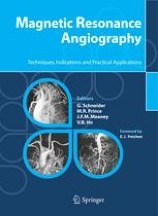
Magnetic resonance angiography : techniques, indications and practical applications
The advent of contrast-enhanced MRA in the early to mid 1990s revolutionized the clinical approach to vascular imaging: an accurate non-invasive imaging modality, not requiring ionizing radiation or potentially nephrotoxic iodinated contrast media, was able to compete with the more hazardous and invasive catheter angiography. Today, MRA is a safe, easy-to-perform procedure routinely used in most imaging centers, and the continued development of faster, more powerful magnets and more effective contrast agents is increasingly helping to overcome many of the early limitations of the technique.
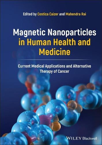
Magnetic nanoparticles in human health and medicine : Current medical applications and alternative therapy of cancer
Progress in improving diagnosis by magnetic resonance imaging (MRI) and using non-invasive and non-toxic magnetic nanoparticles for targeted drug delivery. Focusing on cancer diagnosis and therapy, the book covers both fundamental principles and advanced theoretical and experimental research on the magnetic properties, biocompatibilization, biofunctionalization, and application of magnetic nanoparticles in nanobiotechnology and nanomedicine.
