Refine Results
Sort By
From
To
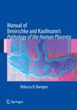
Benirschke and Kaufmann's Pathology of the Human Placenta has long been regarded as the gold standard in the field. It is comprehensive and thorough and contains the detail necessary for those in the subspecialties of placental, perinatal and pediatric pathology. However, placentas are relatively common specimens and are not examined primarily by specialists in the field, but by general pathologists. Thus, there is a need for a more practical and concise manual that can be used by pathology trainees and generalists in their daily work. Manual of Benirschke and Kaufmann's Pathology of the Human Placenta will fill that need. The Manual is a practical, user-friendly guidebook for the general pathologist and pathologist in training for everyday, bench-side use. Organized in 27 chapters, the book will discuss placental development, general features, approach to the specimen by macroscopic and microscopic evaluation, all aspects of placental abnormalities and lesions, disease processes and the placenta, legal aspects of the placental examination, future directions and much more. The sections on macroscopic and microscopic evaluations feature quick-reference tables that allow the reader to identify abnormalities, learn the situations where they occur, and refer back to the text for in-depth discussions. Each chapter will end with selected readings from Pathology of the Human Placenta for more detailed discussions, classic recommended readings, as well as an up-to-date bibliography of current literature. The manual features over 444 illustrations, more than 100 of them in full-color. A must-have for every pathologist and pathology resident.
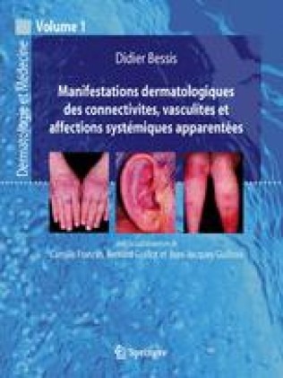
Deals with all the skin and mucous membrane manifestations observed during the various so-called systemic, common or rare diseases. Each chapter covers in an exhaustive and original way the clinical, anatomopathological and therapeutic knowledge essential to the practice of dermatology and internal medicine. The work is abundantly illustrated with color clinical iconography, carefully selected for its clinical representativeness. The didactic anatomopathological figures have been enriched with explanatory diagrams facilitating their reading by the practitioner unfamiliar with dermatological histology. Produced by a group of leading authors in their field, this book is intended for dermatologists, rheumatologists and internists as well as all professionals concerned with systemic diseases and internal medicine.
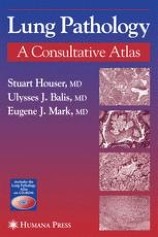
Learning the diagnostic elements of lung pathology requires not only great familiarity with a wide diversity of cases, but also a sharp eye for analyzing pictorial images. In Lung Pathology: A Consultative Atlas, leading experts offer a novel and substantive approach to the teaching of pulmonary pathology. Drawing on 263 challenging, yet exemplary, referral cases taken from files collected over 20 years by internationally renowned lung pathologist, Dr. Eugene Mark, the authors introduce his state-of-the-art approach to the interpretation of pulmonary pathology. This text includes the primary and/or differential diagnosis and the pertinent histological features of each case, as well as clinical history, when available. Key words or phrases in the text are highlighted and digitally hyperlinked to associated images or regions of interest within those images to assist the readers in their correlation. Novel and user friendly, Lung Pathology: A Consultative Atlas describes a cutting-edge diagnostic approach to pulmonary pathology, describing its principles and demonstrating its application in text and full-color illustrations drawn from 263 difficult cases of human lung pathologies.
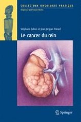
Localized tumors are discovered at an earlier stage. The concept of active surveillance emerges. Minimally invasive treatments such as cryotherapy or radiofrequency for selected cases are now available. The indications for elective open partial nephrectomy are expanding and laparoscopic and then robotic partial nephrectomy are being evaluated.
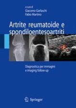
Recent technological evolutions in diagnostic imaging have allowed him to grasp the anatomical and functional alterations that characterize the onset phase of degenerative and inflammatory arthropathies. In the context of the latter, in particular, imaging is not only fundamental in the diagnosis of the early stage of arthritis but also in defining their evolution and response to modern therapeutic treatment. The evaluation of a new imaging semeiology, which makes considerable use of the use of contrast medium in relation to both power Doppler and magnetic resonance imaging, has made it possible to narrow the gap between histology and imaging in rheumatological diagnostics.

I have been involved in the treatment of chronic renal insuf? new and unexpected pathological conditions have also appeared as complications of long-term dialysis. One of these involves polycystic changes and their malignant transformation in diseased kidneys. Since I have studied these polycystic changes and their malignant transformation for many years, I decided to compile the results of my work in a book. Such conditions of diseased kidneys pose serious problems, particularly in Japan, where renal transplantation is performed very infrequently compared with other countries, and a large number of patients are managed by dialysis over a long period.
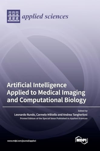
Medical imaging and computational biology continuously pose new fundamental medical and biological questions that often give rise to novel challenges in Artificial Intelligence. These research fields present an increasing need for the application of cutting-edge computational approaches that generally involve machine learning or computational intelligence techniques, which can effectively perform bioimage and biosignal processing in different clinical areas.
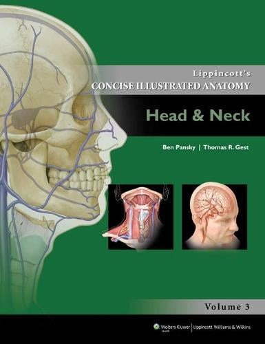
Presents human gross anatomy in an efficient, easy-to-use format by combining core, need-to-know content with detailed atlas-style illustrations. This volume focuses on anatomical structures and functions of the head and neck, and includes clinical considerations concerning these important regions. With artwork adopted from the Lippincott Williams & Wilkins Atlas of Anatomy as well as new illustrations, understanding the functional and clinical relevance of anatomy has never been more at hand!
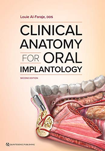
Designed with the practicing implantologist in mind, and it has been revitalized to have the utmost relevance to the clinical reality of oral implantology today. Impeccable full-page illustrations demonstrate a detailed view of each anatomical area, and clinical photos, radiographs, CBCT scans, and cadaver specimens provide a complete picture of what the clinician can expect to encounter.
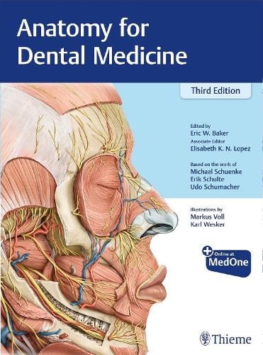
Strikes an optimal balance between systemic and regional approaches to complex head and neck anatomy. Award-winning full-color illustrations, succinct text, summary tables, and questions put anatomical structures and knowledge into a practical context. NEW! Additional radiologic images and landmark features throughoutNEW! Reorganized brain/nerve sectionsNEW! Expanded clinical question appendix including patient box questions in the style of the INBDENEW! Factual question appendix places greater emphasis on areas including the skull, larynx, cross sectional anatomy, body below the neck, and local anesthesia

Daraa Highway - Ghabagheb - Syria
+963-15 2050 - 15 860 380 /1/2/3 - 11 662 3900