


Page 4
Page 4
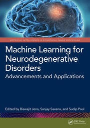
Machine learning for neurodegenerative disorders : advancements and applications
Explores the application of machine learning to the understanding, early diagnosis, and management of neurodegenerative disorders. With a specific focus on its role in ongoing clinical trials, the book covers essential topics such as data collection, pre-processing, feature extraction, model development, and validation techniques. It delves into the applications of neuroimaging techniques like magnetic resonance imaging (MRI), computed tomography (CT), and positron emission tomography (PET) in the diagnosis and understanding of neurodegenerative disorders. Additionally, the book examines various machine-learning algorithms employed for biomarker discovery in neurodegenerative disorders. It highlights the role of neuroinformatics and big data analysis in advancing the understanding and management of neurodegenerative disorders. Furthermore, the book reviews future prospects and presents the ethical considerations and regulatory challenges associated with implementing machine learning approaches in the diagnosis, treatment, and prevention of neurodegenerative disorders.
Liver MRI : Correlation with other Imaging Modalities and Histopathology
In this seminal manuscript the - thor described a new imaging technique which moved the single dimension of NMR spectroscopy to the dual dimension of spatial orientation, thereby resulting in the foundation of modern magnetic re- nance (MR) imaging. Over the ensuing years, MR imaging has assumed an increasingly important role in clinical imaging. It distinguishes itself from other imaging modalities, such as ultrasound (US) or computed tomography (CT), by the unique ability to visualize specific tissue components in a non-- vasive manner. In the earlier days, diagnostic MR imaging was limited to cerebral and musculoskeletal diseases. - aging of other areas which are more prone to movement through breathing (abdominal) or pulsation motions (cardiac) became available more recently, with the introduction of faster sequences and the - velopment of more dedicated MR imaging coils.

Lasers in the Conservation of Artworks ; LACONA VI Proceedings, Vienna, Austria, Sept. 21--25, 2005
In 1995 Professor Costas Fotakis brought together restorers and scientists to discuss the potential of lasers in art conservation. Since then the field of "Lasers in the Conservation of Artworks" has gained enormously in importance. Nowadays restorers and laser scientists work close together in order to develop new fields of applications during the last years.

Clinical MR Imaging : A Practical Approach
This book offers practical guidelines for performing efficient and cost-effective MRI examinations in daily practice. The underlying idea is that, by adopting a practical protocol-based approach, the work-flow in a MRI unit can be streamlined and optimized.
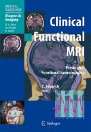
Clinical Functional MRI : Presurgical Functional Neuroimaging
Functional magnetic resonance imaging (fMRI) has contributed significantly to progress in neuroscience by permitting noninvasive imaging of the "human brain at work" under physiological conditions. Within clinical neuroimaging, fMRI is opening up a new diagnostic field by measuring and visualizing brain function. However, fMRI is not yet a standard diagnostic imaging procedure. This textbook is devoted to preoperative fMRI in patients with brain tumors and epilepsies, which are the most well-established clinical applications. By localizing and lateralizing specific brain functions in individual patients, as well as epileptogenic zones, fMRI facilitates the selection of a safe treatment and the planning and performance of function-preserving neurosurgery.
Clinical Cardiac MRI
MRI has become the preferred noninvasive imaging modality for the heart and great vessels. The substantial technological progress achieved in recent years has provided the user with state of the art MRI systems, but their optimal use can be limited by restricted awareness of the potential patient benefit and the necessity for teaching. This extensively illustrated volume, has been specifically compiled to meet these needs. Essential theoretical background information is provided, and imaging acquisition and potential pitfalls are considered in detail. Most importantly, structured guidelines are provided on the interpretation of clinical data in the wide range of cardiac pathology that can be encountered. Throughout, the emphasis is on the implementation of cardiac MRI in clinical practice.
Clinical Blood Pool MR Imaging
MR Angiography covers the basic techniques, safety, efficacy, image processing and pharmaco-economic details to successfully implement a new level of MRA image quality with this new contrast agent.
Classic Papers in Modern Diagnostic Radiology
The subject of diagnostic radiology is now very large and radiology depa- ments are involved in all areas of modern patient care.The defining event in m- ern radiology,and arguably the most significant development in radiology since Wilhelm Röntgen discovered X-rays, was the invention of the CT scanner in the 1970s.The CT scanner introduced modern cross-sectional imaging and also di- tal imaging.We now have MRI and ultrasound and these techniques are replacing many traditional X-ray procedures.The developments in radiology have been the result of a fruitful interaction between the basic sciences, clinical medicine and the manufacturers. This can be seen by looking at the various sources of these publications. Change is produced by the interactions between the various dis- plines. The editors have had a very difficult task in selecting the key discoveries and descriptions.The radiological literature is very large.Medical imaging continues to develop rapidly and these papers are the foundations of our current practice.

Care and Design: Bodies, Buildings, Cities
Connects the study of design with care, and explores how concepts of care may have relevance for the ways in which urban environments are designed. It explores how practices and spaces of care are sustained specifically in urban settings, thereby throwing light on an important arena of care that current work has rarely discussed in detail.
Cardiovascular MRI in practice : A teaching file approach
This text resource outlines the systematic approach to CMR interpretation. The depiction of a "core exam" and the modifications used for a variety of patient circumstances are demonstrated using simple visual assessment of the images. Special emphasis on the advantages of CMR relative to other modalities reinforces practical learning objectives, organized so that the reader starts with patient images – as one would in a clinical scenario – and works back to the didactic material.
Cardiovascular MRI : 150 Multiple Choice Questions and Answers
A Practical Guide for Cardiovascular Magnetic Resonance Imaging provides a comprehensive and reader-friendly educational tool for physicians starting to work with CMR and cardiology and radiology trainees preparing for the Board certification examination.

Cancer du sein ; Compte rendu cours supérieur francophone de cancérologie, (Saint-Paul-de-Vence 13-15 Janvier 2005) = Breast cancer ; Report on the advanced French-language course in oncology, (Saint-Paul-de-Vence, January 13-15, 2005)
In January 2005, the 9th Cours de Saint-Paul-de-Vence, devoted to Breast Cancer, was dominated by the establishment of "Recommendations for Clinical Practice". The five subjects of these recommendations, such as the management of the axillary cavity, neo-adjuvant treatments, the management of patients whose tumor overexpresses HER2, adjuvant hormone therapy and finally adjuvant chemotherapy have been widely discussed. As in each course, certain essential fields were treated such as organized screening, conventional radiology and MRI, prognostic and predictive factors. Ductal cancers and lobular cancers have been compared in a remarkable way in three papers which were produced by the Saint-Cloud school. Data on in situ cancers have been updated. Finally, advice was given for the use of the various standards, whether local, regional or national.R2, l¹hormonothérapie adjuvante et enfin la chimiothérapie adjuvante ont été largement abordés.
Breast MRI : Fundamentals and technical aspects
The text walks the reader through the basics of MRI, making it especially accessible to beginners. From a detailed outline of equipment prerequisites for obtaining high quality breast MRI to instructions on how to optimize image quality, expanded discussions on how to obtain optimized dynamic information.
Breast MRI : Diagnosis and Intervention
This superbly illustrated practical guide is an excellent resource on all aspects of breast MRI for practicing radiologists, oncologists, and surgeons, as well as residents and fellows. Drs. Elizabeth Morris and Laura Liberman, two experts in the field from the Memorial Sloan-Kettering Cancer Center, have collaborated with colleagues from their institution and selected medical centers to share their expertise. Introductory chapters are devoted to diagnosis and cover the basics of performing breast MRI exams, setting up a breast MRI program, and understanding clinical indications. Additional chapters discuss breast interventional procedures including MRI-guided needle localization, MRI-guided biopsy, and percutaneous ablation of breast cancer; MRI of breast implants.

Breast cancer ; 2nd ed.
This book highlights M. D. Anderson Cancer Center’s multidisciplinary approach and reviews the entire spectrum of patient care, from prevention and screening through diagnosis and treatment through posttreatment follow-up and survivorship issues.
Bone densitometry for technologists
Radiologic technologists, nurse practitioners, physician assistants, and dedicated densitometry technologists can find new guidelines for bone density testing, new therapies for osteoporosis, and new treatment guidelines for osteoporosis, as well as new chapters on pediatric densitometry body composition assessments, and the use of skeletal morphometry in diagnosis and fracture risk prediction. The twelve appendices have also been updated to reflect the most current information available, including contacts for densitometry equipment manufacturers and organizations, guidelines for bone density testing and CPT codes, definitions of terms, and new conversion equations.

Bioreactors for Tissue Engineering : Principles, Design and Operation
This volume addresses the issue of mechanical conditioning of the tissue, and describes the use of techniques such as MRI for monitoring tissue growth. It also deals with the application of bioreactor technology to tissue engineering products.

Atlas of topographical and pathotopographical anatomy of the head and neck
Presents the ultrasonic topographical and pathotopographical anatomy of the head and neck, offering further detail into these important areas for use by medical professionals. This atlas of topographic and pathotopographic human anatomy is a fundamental and practically important book designed for doctors of all specializations and students of medical schools. Here you can find almost everything that is connected with the topographic and pathotopographic human anatomy, including original graphs of logical structures of topographic anatomy and development of congenital abnormalities, topography of different areas in layers, pathotopography, computer and magnetic resonance imaging (MRI) of topographic and pathotopographic anatomy.

Atlas of Regional Anatomy of the Brain Using MRI : with functional correlations
The volume provides a unique review of the essential topographical anatomy of the brain from an MRI perspective, correlating high-quality anatomical plates with the corresponding high-resolution MRI images. The book includes a historical review of brain mapping and an analysis of the essential reference planes used for the study of the human brain. Subsequent chapters provide a detailed review of the sulcal and the gyral anatomy of the human cortex, guiding the reader through an interpretation of the individual brain atlas provided by high-resolution MRI.

Atlas of oral and maxillofacial radiology
Presents an extensive case collection of both common and less common conditions of the jaws and teeth. Focusing on the essentials of radiologic interpretation, this is a go-to companion for clinicians in everyday practice who have radiologically identified a potential abnormality, as well as a comprehensive study guide for students at all levels of dentistry, surgery and radiology. Unique lesion-based problem solving chapter makes this an easy-to-use reference in a clinical settingIncludes 2D intraoral radiography, the panoramic radiograph, cone beam CT, multidetector CT and MRIMultiple cases are presented in order to demonstrate the variation in the radiological appearances of conditions affecting the jaws and teethSpecial focus on conditions where diagnostic imaging may substantially contribute to diagnosisFeatures a useful chapter covering the temporomandibular joint
