


Page 2
Page 2

Medical Imaging and Augmented Reality ; 3rd International Workshop, Shanghai, China, August 17-18, 2006, Proceedings
The Third International Workshop on Medical Imaging and Augmented Reality, MIAR 2006, was held in Shanghai, China at the Regal International East Asia Hotel during August 17-18, 2006. The goal of MIAR 2006 was to bring together researchers in medical image computing and intervention to present the state-of-the-art devel- ments in this ever-growing research area.

Maxillofacial Imaging
Maxillofacial imaging has evolved dramatically over the past two decades with development of new cross-sectional imaging techniques. Traditional maxillofacial imaging was based on plain films and dental imaging. However, today’s advanced imaging techniques with CT and MRI have only been partially implemented for maxillofacial questions. This book bridges the gap between traditional maxillofacial imaging and advanced medical imaging. We have applied CT and MRI to a variety of maxillofacial cases and these are illustrated with high-quality images and multiple planes. A comprehensive chapter on imaging anatomy is also included. This book is useful for oral and maxillofacial radiologists, oral and maxillofacial surgeons, dentists, radiologists, plastic surgeons, head and neck surgeons, and others that work with severe maxillofacial disorders.

Maxillofacial Imaging
Demonstrates how advanced medical imaging techniques can be successfully applied to dental and maxillofacial conditions. There is a focus on CT and MRI, but the use of all contemporary imaging techniques are illustrated including PET, PET/CT, ultrasonography, and cone beam CT. The presentation is in atlas style, with succinct, bulleted text and a wealth of high-quality images in multiple planes. All images for each patient are grouped to enable the reader very quickly to gain an imaging overview of the condition under consideration. After a comprehensive introductory chapter on normal imaging anatomy, the role of advanced imaging techniques is described in pathologic conditions of the mandible and maxilla, temporomandibular joint, regions closely related to the jaw, paranasal sinuses, oral cavity, salivary glands, and structures adjacent to the maxillofacial region. A concluding chapter examines the use of interventional procedures for diagnosis and treatment of maxillofacial conditions. Compared with the first edition, numerous additional cases have been incorporated and a completely new chapter focuses on cone beam CT.

Inverse Problems and Imaging : Lectures given at the C.I.M.E. Summer School held in Martina Franca, Italy September 15–21, 2002
Nowadays we are facing numerous and important imaging problems: nondestructive testing of materials, monitoring of industrial processes, enhancement of oil production by efficient reservoir characterization, emerging developments in noninvasive imaging techniques for medical purposes - computerized tomography (CT), magnetic resonance imaging (MRI), positron emission tomography (PET), X-ray and ultrasound tomography, etc. In the CIME Summer School on Imaging (Martina Franca, Italy 2002), leading experts in mathematical techniques and applications presented broad and useful introductions for non-experts and practitioners alike to many aspects of this exciting field. The volume contains part of the above lectures completed and updated by additional contributions on other related topics: a general presentation and introduction (Moscoso), X-ray tomography (Natterer), Electromagnetic imaging (Dorn, Bertete-Aguirre, Papanicolaou), coherent imaging in telecommunications in a multiple input-multiple output setup (Dorn), polarization based optical imaging (Moscoso), topological derivatives used in shape reconstruction related to inverse scattering problems (Carpio, Rapún), Point interactions (Dell’Antonio, Figari, Teta).

Information Processing in Medical Imaging ; 20th International Conference, IPMI 2007, Kerkrade, The Netherlands, July 2-6, 2007, Proceedings
The 20th International Conference on Information Processing in Medical Im- ing(IPMI)washeldduringJuly2–6,2007,atRolducAbbey,locatedinKerkrade in the south of the Netherlands. IPMI is one of the longest running conferences in medical imaging.
Information processing in medical imaging ; 19th International conference, IPMI 2005, Glenwood Springs, CO, USA, July 10-15, 2005, Proceedings
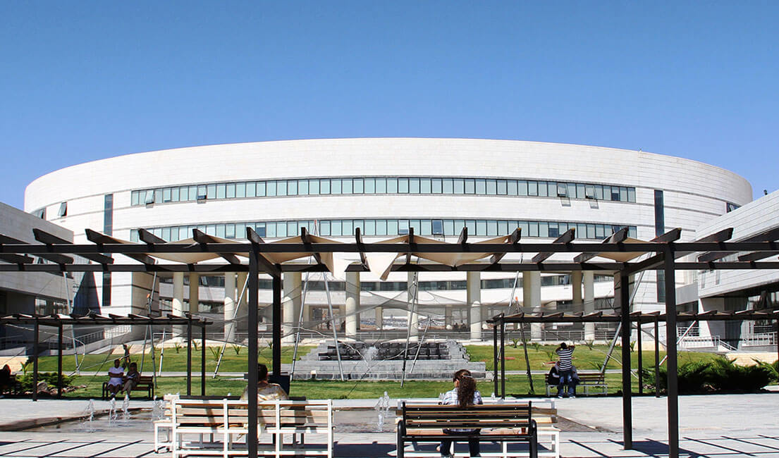
The nineteenth biennial International Conference on Information Processing in Medical Imaging (IPMI) was held July 11–15, 2005 in Glenwood Springs, CO, USA on the Spring Valley campus of the Colorado Mountain College. Following the successful meeting in beautiful Ambleside in England, this year’s conference addressed important recent developments in a broad range of topics related to the acquisition, analysis and application of biomedical images. Interest in IPMI has been steadily growing over the last decade. This is p- tially due to the increased number of researchers entering the ?eld of medical imagingasaresultoftheWhitakerFoundationandtherecentlyformedNational Institute of Biomedical Imaging and Bioengineering. This year, there were 245 full manuscripts submitted to the conference which was twice the number s- mitted in 2003 and almost four times the number of submissions in 2001. Of these papers, 27 were accepted as oral presentations, and 36 excellent subm- sions that could not be accommodated as oral presentations were presented as posters. Selection of the papers for presentation was a di?cult task as we were unable to accommodate many of the excellent papers submitted this year. All accepted manuscripts were allocated 12 pages in these proceedings.

Imaging Pelvic Floor Disorders
This volume builds on the success of the first edition of Imaging Pelvic Floor Disorders and is aimed at those practitioners with an interest in the imaging, diagnosis and treatment of pelvic floor dysfunction. Concise textual information from acknowledged experts is complemented by high-quality diagrams and images to provide a thorough update of this rapidly evolving field. Introductory chapters fully elucidate the anatomical basis underlying disorders of the pelvic floor. State of the art imaging techniques and their application in pelvic floor dysfunction are then discussed in detail. Additions since the first edition include consideration of the effect of aging and new chapters on perineal ultrasound, functional MRI and MRI of the levator muscles.

Imaging of the Shoulder : Techniques and Applications
This volume covers the broad spectrum of imaging methods and abnormalities of relevance in the diagnostic workup of the shoulder. In the first part of the book, individual chapters are devoted to radiography, arthrography, computed tomography and CT arthrography, magnetic resonance imaging and MR arthrography, ultrasound and interventional procedures. The second part of the book then documents the application of these techniques to each of the clinical problems and diseases encountered in the shoulder. The authors are all experts in their field and include rising stars of musculoskeletal radiology.

Imaging of Soft Tissue Tumors
This book provides a comprehensive survey of the growing role of medical imaging studies in the detection, staging, grading, tissue characterization, and post-treatment follow-up of soft tissue tumors. For each tumor group, imaging findings are correlated with clinical, epidemiologic, and histologic data.

Imaging of Orbital and Visual Pathway Pathology
This is one of the first books to deal with imaging of pathology of the entire visual system. It is divided into two parts, general and special. In the general part, the most important basics of modern imaging methods are discussed in detail, but with less emphasis on the physical background than in purely neuro-/radiological books. A chapter is devoted to the meticulous presentation of imaging anatomy of the orbit and intracranial visual pathway.

Imaging Atlas of the Pelvic Floor and Anorectal Diseases
The goal of Imaging Atlas of the Pelvic Floor and Anorectal Diseases is to clearly and precisely present indications, techniques, limitations, sources of errors, and pitfalls of these imaging modalities.
Image-Guided Interventions : Technology and Applications
Responding to the growing demand for minimally invasive procedures, Image-Guided Interventions: Technology and Applications provides a cohesive overview of the current technological advances in image-guided surgery, and their applications in the clinical environment.

Image-Guided IMRT
Intensity-modulated radiation therapy (IMRT), one of the most important developments in radiation oncology in the past 25 years, involves technology to deliver radiation to tumors in the right location, quantity and time. Unavoidable irradiation of surrounding normal tissues is distributed so as to preserve their function. The achievements and future directions in the field are grouped in the three sections of the book, each suitable for supporting a teaching course.

Image Processing in Radiology : Current Applications
Few fields have witnessed such impressive advances as image processing in radiology. The progress achieved has revolutionized diagnosis and greatly facilitated treatment selection and accurate planning of procedures. This book provides a comprehensive and up-to-date description of how to use 2D and 3D processing tools in clinical radiology.

How does MRI work? : An introduction to the physics and function of magnetic resonance imaging
A succinct introduction to the physics and function of magnetic resonance imaging with an emphasis on practical information. It progresses to more advanced techniques such as parallel imaging and cardiovascular MR imaging.

High-Grade Gliomas : Diagnosis and Treatment
This is truly an exciting time in the field of neuro-oncology, particularly in the area of hi- grade gliomas. The management of patients with high-grade gliomas has historically been one of the most challenging and disheartening fields in medicine, where failure is the rule and longevity is the exception. The jaded often state that despite purported advances in surgical and radiotherapeutic techniques and a myriad of clinical trials of medical therapies, the s- vival statistics for glioblastoma have not changed in the last three decades. The nihilism associated with these tumors is such that some practitioners still advise against treatment or even biopsy, recommending palliative care with the diagnosis based only on history and an MRI scan. If the current state-of-the-art in the diagnosis and management of high-grade gliomas was truly so bleak, there would be no reason to compile and publish a monograph on the subject. The fact is that we have recently entered an era where real progress is being made in our understanding and treatment of high-grade gliomas that is directly benefiting some patients. We are slowly but surely chipping away at this problem. One approach has exploited correlations between particular molecular markers and therapeutic response.

High Field Brain MRI : Use in Clinical Practice
This book describes the development of systems of magnetic resonance imaging using the higher magnetic field strength of 3 tesla, in comparison to the current gold standard of 1.5 tesla. It is the first monography on high-field Brain MR in Neuroradiology

Handbook of high field dynamic nuclear polarization
This comprehensive handbook is a compendium of the current state-of-the art of high field Dynamic Nuclear Polarization—from long-proven, early developments, up to today’s hot topics. It covers all the relevant subjects that have made a direct or indirect contribution toward advancing this field, and focuses on topics such as: the theory behind the effects seen within DNP; instrumentation required for carrying out DNP; and specific applications of DNP including protein monitoring, catalysis, nanoparticles, biological and clinical studies.

Gastrointestinal Tract Sonography in Fetus and in Children
Sonography of the gastrointestinal tract in fetuses, neonates and children entails no known biological risk, permits serial scanning and can provide information unobtainable with any other imaging modality. In experienced hands it can be used as the initial imaging technique in a number of gastrointestinal diseases and conditions. This book provides a comprehensive account of the current state of the art regarding sonography in this context.

Functional MRI : Basic principles and clinical applications
This practical and timely volume presents an overview of the principles of fMRI and explains key concepts, including scanning methodologies, experimental research design, and data analysis. Informative discussions cover the technical and clinical challenges associated with fMRI. A neuroanatomical atlas adds to the sensible approach and serves as a useful reference by clearly illustrating normal functioning for important areas of the brain.
