


الصفحة 1
الصفحة 1
Mezzi di contrasto in ecografia : Applicazioni addominali = Contrast media in ultrasound : Abdominal applications
Lopera presenta unanalisi completa delle applicazioni dei mezzi di contrasto ecografici a livello addominale. La parte introduttiva di natura tecnica presenta la classificazione generale e le proprietà fisiche dei mezzi di contrasto ecografici, nonché una dettagliata classificazione delle tecniche ecografiche contrasto-specifiche; segue una parte speciale che descrive le applicazioni delle microbolle a livello del fegato, della milza e del rene. Vengono quindi presentate le applicazioni speciali e innovative dei mezzi di contrasto ecografici nellambito dei traumi delladdome, della patologia del pancreas, della parete intestinale e dei vasi addominali, e della valutazione del reflusso vescico-ureterale. Infine, viene proposta unanalisi economica relativa allapplicazione dei mezzi di contrasto ecografici nella caratterizzazione delle lesioni focali epatiche. Il libro è principalmente rivolto ai medici impegnati nellattività ecografica quotidiana, particolarmente in ambito internistico, che desiderano ampliare le proprie competenze nel settore dellecografia con mezzo di contrasto

Medicina delletà prenatale : Prevenzione, diagnosi e terapia dei difetti congeniti e delle principali patologie gravidiche = Prenatal medicine: Prevention, diagnosis and therapy of congenital defects and the main pathologies of pregnancy
This text aims to respond to the actual need to bring together in a single and updated reference text notions and data from different backgrounds, all essential for optimal monitoring of pregnancy.

Ileo meccanico dell’intestino tenue : Aspetti TC e correlazioni eco-radiografiche = Mechanical ileus of the small intestine : CT aspects and echo-radiographic correlations
Mechanical occlusion of the small intestine is a frequently encountered clinical problem, whose clinical-instrumental management is made difficult by the complexity of the mechanisms responsible for the occlusive picture. The role of the radiologist is to promptly guide the therapeutic process on the basis of a correct interpretation of the iconographic pictures. The purpose of this volume - the only one of its kind - is to relate the pathophysiology of occlusion to imaging. The rich iconography, which ranges from clinical onset to resolution, emphasizes the role of multidetector computed tomography with intravenous contrast, which is the current gold standard. However, the correlations with the echo-radiographic methods are not forgotten, which retain an effective diagnostic role, with a low biological and economic cost. The mechanical ileus of the small intestine is a striking example in support of the dream of many professionals: that, that is, to have a figure of a fine abdominal emergency radiologist, an expert in organ and disease and not only in the machine, as often unfortunately it happens.
Fondamenti di ingegneria clinica ; Vol.2 : Ecotomografia = Fundamentals of clinical engineering ; Vol.2 : Ultrasound
With the second volume of Clinical Engineering, the Author intends to provide an updated scientific-didactic overview of the physical principles of ultrasound, of the technology and of the implementation aspects of the ecotomograph.

Ecografia in oncologia : Testo-Atlante di ultrasonologia diagnostica e interventistica dei tumori = Ultrasound in oncology : Text-Atlas of diagnostic and interventional ultrasound of tumors
Offers detailed information on the key applications of different ultrasound techniques to the study of tumors. The necessary background on the major problems of clinical oncology is provided: screening, identification, characterization and staging of malignant neoplasms, evaluation of the therapeutic response, follow-up. The examination technique and the interpretative principles of all ultrasound techniques are carefully discussed (grayscale ultrasound, color-Doppler, pulsed Doppler, power-Doppler, echocontrastography), with particular attention to the practical aspects, to the "tricks of the trade. "and the potential methodological and semeiological pitfalls. Particular emphasis is placed on those areas where ultrasound plays a decisive role compared to other diagnostic imaging methods, with a practical and original approach, for "clinical problems". The main eco-interventional, diagnostic and therapeutic procedures are illustrated in detail. The very rich iconography, also including a CD-ROM with original videos, punctually integrates the text. A compact and homogeneous work, necessary for all doctors, both radiologists and non-radiologists, who deal with ultrasound.

Ecografia e procedure interventistiche percutanee: Fegato, vie biliari e pancreas = Ultrasound and percutaneous interventional procedures : Liver, bile ducts and pancreas
The volume presents a concise overview of the most common percutaneous interventional procedures and is intended as a quick guide for the radiologist and sonographer who, more and more frequently, are required to perform interventional procedures on the liver, biliary tract and pancreas for both diagnostic and therapeutic.

Ecografia dellapparato osteoarticolare : Anatomia, semeiotica e quadri patologici = Ultrasound of the osteoarticular system: Anatomy, semeiotics and pathological pictures
The echography is useful in the valutazione of articolari pathology in peace with rheumatic malattia and, other than in the diagnosis, is effective in the monitoring with power Doppler of the synovite, with a higher sensitivity to all'esame clinical. The high resolution ultrasound, integrated with the power Doppler technique, even to the degree of individualizing the entheopathic anomaly, the osseous erosion and the tendine involvement. The objective of the operation is to present the technique of same, and progress of echography in muscoloscheletric rheumatic pathology and the diagnosis of ultrasound in articolare rheumatic pathology. L’iconography è arricchita with integrated imaging of ultrasound, conventional radiology, CT and MRI.
Malattia coronarica : Fisiopatologia e diagnostica non invasiva con TC = Coronary heart disease: Pathophysiology and non-invasive diagnostics with CT
The work stems from the desire to provide the cardiologist or non-imaging physician with the basis for understanding technical principles and methods of acquiring and reconstructing images. At the same time, even the radiologist who does not have specific experience in cardiac imaging will be able to acquire basic knowledge of coronary anatomy and pathophysiology.
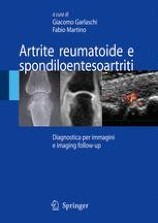
Artrite reumatoide e spondiloentesoartriti : Diagnostica per immagini e imaging follow-up = Rheumatoid Arthritis and Spondyloentesoarthritis : Follow-up imaging and imaging
Recent technological evolutions in diagnostic imaging have allowed him to grasp the anatomical and functional alterations that characterize the onset phase of degenerative and inflammatory arthropathies. In the context of the latter, in particular, imaging is not only fundamental in the diagnosis of the early stage of arthritis but also in defining their evolution and response to modern therapeutic treatment. The evaluation of a new imaging semeiology, which makes considerable use of the use of contrast medium in relation to both power Doppler and magnetic resonance imaging, has made it possible to narrow the gap between histology and imaging in rheumatological diagnostics.
