
الصفحة 139
الصفحة 139
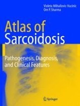
Atlas of Sarcoidosis : Pathogenesis, Diagnosis and Clinical Features
An Atlas of Sarcoidosis: Pathogenesis, Diagnosis and Clinical Features combines illustrations and clinical images of the authors' extensive practices, so that readers have unparalleled access to a comprehensive collection of sarcoidosis images. The atlas is designed to complement and provide a visual supplement to already existing texts on sarcoidosis. Each organ involvement is dealt in a brief and easy to comprehend manner. Various radiographic and laboratory abnormalities are then linked to the clinical features in order to encourage a smooth and easy practical integration at the bedside and to help practising pulmonologists, dermatologists and other clinicians who require a comprehensive visual encyclopedia of sarcoidosis images.
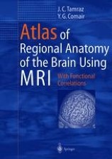
Atlas of Regional Anatomy of the Brain Using MRI : with functional correlations
The volume provides a unique review of the essential topographical anatomy of the brain from an MRI perspective, correlating high-quality anatomical plates with the corresponding high-resolution MRI images. The book includes a historical review of brain mapping and an analysis of the essential reference planes used for the study of the human brain. Subsequent chapters provide a detailed review of the sulcal and the gyral anatomy of the human cortex, guiding the reader through an interpretation of the individual brain atlas provided by high-resolution MRI.

Atlas of Psoriatic Arthritis
A visual guide to the complex and inter-related diseases of psoriatic arthritis and psoriasis, this volume contains more than 150 images spanning a wide spectrum of variations of the disease.

Atlas of Practical Applications of Cardiovascular Magnetic Resonance
The Atlas of Practical Applications of Cardiovascular Magnetic Resonance contains over two hundred illustrations and a Glossary of terms. This atlas will assist cardiologists to determine when a CMR exam is useful for diagnosis and provide details on how to plan and read CMR studies.
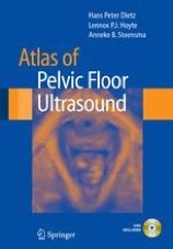
Atlas of Pelvic Floor Ultrasound
Atlas of Pelvic Floor Ultrasound provides an introduction to pelvic floor imaging, as well as a resource to be used during initial and more advanced practice.
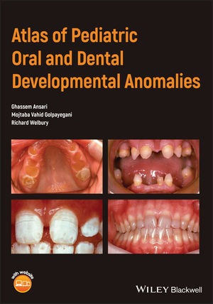
Atlas of Pediatric Oral and Dental Developmental Anomalies
A quick reference diagnostic guide for students and clinicians, covering a wide range of oral and dental developmental anomalies in children and adolescents. A useful, quick reference atlas helping students and clinicians diagnose a wide range of oral and dental developmental anomalies in children and adolescents Highly illustrated with clinical photographs Describes both common and rare conditions, and explores treatment options

Atlas of orthodontic case reviews
Offers a comprehensive resource to the treatment of orthodontic malocclusions with a case-based approach. Discusses and illustrates the treatment of orthodontic malocclusions using actual clinical casesPresents more than 800 clinical photographs showing the stages of each treatment, to act as a visual referenceIncludes a description of each malocclusion, an explanation of the desired treatment outcomes, an account of the changes, and review questions for each case

Atlas of Organ Transplantation
A comprehensive compilation of the majority of surgical procedures in transplant surgery, this book details the latest and most innovative procedures in one reference work. Atlas of Organ Transplantation is essential reading for all transplant surgeons, residents and fellows, as well as operating room nurses and transplant nurse coordinators.
Atlas of oral and maxillofacial surgery
Enhances your surgical skills with Atlas of Oral and Maxillofacial Surgery. It offers detailed, step-by-step instructions and more than 2,500 full-color illustrations that demonstrate how to plan for and perform oral and maxillofacial surgical procedures safely and efficiently. Comprehensive and expanded coverage addresses the broad scope of the specialty, ranging from the surgical anatomy of the head and neck to oral surgery, implant surgery, orthognathic and craniofacial surgery, cleft lip and palate, craniomaxillofacial trauma, head and neck oncology, reconstructive procedures, TMJ surgery, facial cosmetic surgery, obstructive sleep apnea, and more.

Atlas of oral and maxillofacial radiology
Presents an extensive case collection of both common and less common conditions of the jaws and teeth. Focusing on the essentials of radiologic interpretation, this is a go-to companion for clinicians in everyday practice who have radiologically identified a potential abnormality, as well as a comprehensive study guide for students at all levels of dentistry, surgery and radiology. Unique lesion-based problem solving chapter makes this an easy-to-use reference in a clinical settingIncludes 2D intraoral radiography, the panoramic radiograph, cone beam CT, multidetector CT and MRIMultiple cases are presented in order to demonstrate the variation in the radiological appearances of conditions affecting the jaws and teethSpecial focus on conditions where diagnostic imaging may substantially contribute to diagnosisFeatures a useful chapter covering the temporomandibular joint
Atlas of oral and maxillofacial anatomy
Enables readers to observe the anatomy from the same view as seen during invasive clinical procedures. This is critical for a better understanding of these procedures, and surgical annotations are included as necessary. Atlas of Oral and Maxillofacial Anatomy is the first book of its kind to be devoted to the clinical anatomy of the region for dentists and oral and maxillofacial surgeons. It will satisfy the demand for such a comprehensive atlas in this field of surgery and will be welcome and timely for clinicians and trainees. Beyond specialists and residents in oral and maxillofacial surgery and general dentists, the book will be of value for craniofacial surgeons, anatomists, plastic surgeons, ENT surgeons, head and neck surgeons, neurosurgeons, dental students, medical students, dental hygienists, and nurses working with dentists and oral and maxillofacial surgeons. Top quality photographs of fresh cadaveric dissections / Anatomy shown as seen during invasive clinical procedures / Ideal aid for dentists and oral and maxillofacial surgeons
Atlas of Operative Oral and Maxillofacial Surgery
Comprised of concise text and detailed vignettes focusing on surgical indications, contraindications, pertinent anatomy, virtual surgical planning, operative techniques, postoperative management, complications and key points with over 2,000 high-quality images. Serves as an innovative, multidisciplinary, surgical atlas covering core aspects of oral and maxillofacial surgery, head and neck reconstructive surgery, and facial cosmetic surgery. Chapters are written by experts in their fields and are designed to provide high-yield information utilizing a case report format.
Atlas of Non-Invasive Coronary Angiography by Multidetector Computed Tomography
The multidetector CT scanner speeds diagnosis and treatment of patients. One of its many uses is to perform CT coronary angiography. Multidetector CT has generated excitement within the cardiology and radiology community as it provides clear pictures and takes less time than other non-invasive techniques, including conventional spiral and electron-beam CT which can take up to an hour or more. This atlas presents over 160 illustrations, with 116 in color and illustrates the capacity of multidetector CT for the analysis of the anatomy of the coronary arteries.
Atlas of Neuromuscular Diseases : A Practical Guideline
A comprehensive outline of neuromuscular diseases, written by experienced American and European authors. It discusses all aspects of neuromuscular disorders including the cranial nerves, spinal nerves, motor neurone disease, the nerve plexus, peripheral nerves, mononeuropathies, entrapment syndromes, polyneuropathies, the neuromuscular junction, and muscle disease. Each chapter is uniformly structured into anatomy, symptoms, signs, pathogentic possibilities, diagnosis and differential diagnosis, therapy and prognosis. Additionally the diagnostic tools and investigations used in neuromuscular disease are explained and a practical guide is given how to advance from symptoms to syndromes. For each disease the therapeutic options are described. It contains large number of clinical and histologic pictures from the practical experience of the authors and also a number of artists drawings to facilitate the understanding of anatomic structures.
Atlas of Morphology and Functional Anatomy of the Brain
Divided into a morphological and a functional imaging section. The morphological atlas includes 3D surface images, axial, coronal, and sagittal scans acquired with high-definition T2 fast spin echo (FSE) sequences, and standard and inverted-contrast images. The MR scans are shown side by side with the corresponding anatomical brain sections, provided by Prof. Henri Duvernoy, for more effective comparison. The anatomical nomenclature adopted for both the MR and the anatomical images is listed in an jacket flap for easier consultation.

Atlas of mineral deposits distribution in China
Includes instruction of national mineral database 2020 and atlas of national mineral deposits distribution derived from national mineral database 2020. National mineral database 2020 is based on data from National Geological Archives China(NGAC). Moreover, it introduces the construction method and updates maintenance mechanism of the mineral deposits database and proposes the concept of updating data based on collected archives. The construction guideline on national mineral deposits database provides guiding framework for the future development on geological database.
Atlas of mandibular and maxillary reconstruction with the fibula flap : A step-by-step approach
Deals with the standard technique used for reconstructing the mandible and the maxilla - the fibula flap. The reader will find useful information on all issues that are important in the surgical procedure, including the use of CAD-CAM technology (Computer Assisted Technology), bone synthesis and flap modelling. The editors draw on their 30 years of experience to provide a step-by-step description of this surgical procedure. With the help of numerous illustrations, the reader will learn the technical, functional and aesthetic developments since 1989 when this technique was first described.
Atlas of lacrimal surgery
International experts provide a stepwise analysis and describe the clinical management of these patients with detailed medical and surgical treatment plans. Further, the accompanying DVD presents video clips on surgical methods such as endonasal and external DCR; endoscopic versus microscopic procedure; lacrimal endoscopy and transcanalicular surgery; laser DCR and DCR in children; traumatic lesions; interventional radiology and stenting; and conjunctivorhinostomy and Jones tube. With this excellent guide the reader will be able to solve any problem that may occur with the reconstruction of a disturbed or damaged lacrimal system.
Atlas of immediate dental implant loading
Offers an up-to-date and comprehensive overview of the immediate restoration of teeth and immediate functional loading when using different implant systems and surfaces in patients with single tooth loss or partial or complete edentulism.
Atlas of global change risk of population and economic systems
Iillustrates the spatial distribution of the global change risk of population and economic systems with the maps of environment, global climate change, global population and economic systems, and global change risk. The risks of global change are mapped at 0.25 degree grid unit. The risk results and their contribution rates of the world at national level are unprecedentedly derived and ranked. The book can be a good reference for researchers and students in the field of global climate change and natural disaster risk management, as well as risk managers and enterpriser to understand the global change risk of population and economic systems.
