


الصفحة 11
الصفحة 11

Capital Punishment and the Criminal Corpse in Scotland, 1740–1834
This book provides the most in-depth study of capital punishment in Scotland between the mid-eighteenth and early nineteenth century to date. Based upon an extensive gathering and analysis of previously untapped resources, it takes the reader on a journey from the courtrooms of Scotland to the theatre of the gallows. It introduces them to several of the malefactors who faced the hangman’s noose and explores the traditional hallmarks of the spectacle of the scaffold. It demonstrates that the period between 1740 and 1834 was one of discussion, debate and fundamental change in the use of the death sentence and how it was staged in practice. In addition, the study provides an innovative investigation of the post-mortem punishment of the criminal corpse. It offers the reader an insight into the scene at the foot of the gibbets from which criminal bodies were displayed, and around the dissection tables of Scotland’s main universities where criminal bodies were used as cadavers for anatomical demonstration. In doing so it reveals an intermediate stage in the long-term disappearance of public bodily punishment.

Cancer Metastasis and the Lymphovascular System : Basis for Rational Therapy
Sentinel lymph node (SLN) procedures have opened a window of opportunity for the study of micrometastasis. In eighty percent (80%) of metastasis there lies an orderly pattern of progression via the lymphatic network, while 20% of the time systemic metastasis occurs, bypassing the lymphatic system. During the past two decades, significant progress has been achieved in understanding the anatomical, functional, cellular and molecular aspects of the lymphovascular system and the metastasis process.Molecular imaging advances help to localize early cancers more precisely.
Brain and Human Body Modeling 2020 : Computational Human Models Presented at EMBC 2019 and the BRAIN Initiative® 2019 Meeting
This book describes modern applications of computational human modeling in an effort to advance neurology, cancer treatment, and radio-frequency studies including regulatory, safety, and wireless communication fields.
Brain and Human Body Modeling : Computational Human Modeling at EMBC 2018
This book describes modern applications of computational human modeling with specific emphasis in the areas of neurology and neuroelectromagnetics, depression and cancer treatments, radio-frequency studies and wireless communications. Special consideration is also given to the use of human modeling to the computational assessment of relevant regulatory and safety requirements. Readers working on applications that may expose human subjects to electromagnetic radiation will benefit from this book’s coverage of the latest developments in computational modelling and human phantom development to assess a given technology’s safety and efficacy in a timely manner.

Bone augmentation by anatomical region : Techniques and decision-making
It's a unique, evidence-based guide focusing on each specific anatomical region – anterior maxilla, posterior maxilla, anterior mandible, and posterior mandible – in order to emphasize the correct implemented procedures needed to successfully perform oral osseous reconstruction.

Biomedical Simulation ; 3rd International Symposium, ISBMS 2006, Zurich, Switzerland, July 10-11, 2006, Proceedings
This book contains the written contributions to the Third International Sym- sium on Biomedical Simulation (ISBMS), which was held in Zurich, Switzerland, on July 10-11, 2006. The manuscripts are organized around three thematic sections which cover several of the major aspects of our rapidly growing ?eld: anatomical modeling and tissue properties, simulation of biophysical processes, as well as systems and applications. The symposium provided an international forum for researchers interested in using biomedical simulation technology for the improvement of patient care. It was held in the spirit and continuation of the symposia on Surgical Simulation and Soft Tissue Modeling (IS4TM) organized in 2003 by INRIA, and on Medical Simulation (ISMS) in 2004 by CIMIT.

Atoms, molecules and photons : An introduction to atomic- molecular- and quantum physics
This introduction to Atomic and Molecular Physics explains how our present model of atoms and molecules has been developed over the last two centuries both by many experimental discoveries and, from the theoretical side, by the introduction of quantum physics to the adequate description of micro-particles. It illustrates the wave model of particles by many examples and shows the limits of classical description. The interaction of electromagnetic radiation with atoms and molecules and its potential for spectroscopy is outlined in more detail and in particular lasers as modern spectroscopic tools are discussed more thoroughly. Many examples and problems with solutions are offered to encourage readers to actively engage in experimentation.
Atomistic modeling of materials failure
Atomistic Modeling of Materials Failure is an introduction to molecular and atomistic modeling techniques applied to solid deformation and fracture. Focusing on a variety of brittle, ductile and geometrically confined materials, this detailed overview includes computational methods at the atomic scale, and describes how these techniques can be used to model the dynamics of cracks, dislocations and other deformation mechanisms.

Atomic structure theory : Lectures on atomic physics
This is a textbook for students with a background in quantum mechanics. The text is designed to give hands-on experience with atomic structure calculations. Material covered includes angular momentum methods, the central field Schrödinger and Dirac equations, Hartree-Fock and Dirac-Hartree-Fock equations, multiplet structure, hyperfine structure, the isotope shift, dipole and multipole transitions, basic many-body perturbation theory, configuration interaction, and correlation corrections to matrix elements.

Atomic spectroscopy : Introduction to the theory of hyperfine structure
Atomic Spectroscopy provides a comprehensive discussion on the general approach to the theory of atomic spectra, based on the use of the Lagrangian canonical formalism. This approach is developed and applied to explain the hydrogenic hyperfine structure associated with the nucleus motion, its finite mass, and spin. The non-relativistic or relativistic, spin or spin-free particle approximations can be used as a starting point of general approach. The special attention is paid to the theory of Lamb shift formation. The formulae for hydrogenic spectrum including the account of Lamb shift are written in simple analytical form. The book is of interest to specialists, graduate and postgraduate students, who are involved into the experimental and theoretical research in the field of modern atomic spectroscopy.
Atomic Force Microscopy, Scanning Nearfield Optical Microscopy and Nanoscratching : Application to Rough and Natural Surfaces
Making a clear distinction is made between nano- and micro-mechanical testing for physical reasons, this monograph describes the basics and applications of the supermicroscopies AFM and SNOM, and of the nanomechanical testing on rough and technical natural surfaces in the submicron range down to a lateral resolution of a few nm. New or improved instrumentation, new physical laws and unforeseen new applications in all branches of natural sciences (around physics, chemistry, mineralogy, materials science, biology and medicine) and nanotechnology are covered as well as the sources for pitfalls and errors.

Atomic and Nuclear Analytical Methods : XRF, Mössbauer, XPS, NAA and Ion-Beam Spectroscopic Techniques
This book is a blend of analytical methods based on the phenomenon of atomic and nuclear physics. It comprises comprehensive presentations about X-ray Fluorescence (XRF), Mössbauer Spectroscopy (MS), X-ray Photoelectron Spectroscopy (XPS), Neutron- Activation Analysis (NAA), Particle Induced X-ray Emission Analysis (PIXE), Rutherford Backscattering Analysis (RBS), Elastic Recoil Detection (ERD), Nuclear Reaction Analysis (NRA), Particle Induced Gamma-ray Emission Analysis (PIGE), and Accelerator Mass Spectrometry (AMS). These techniques are commonly applied in the fields of medicine, biology, environmental studies, archaeology or geology et al. and pursued in major international research laboratories.
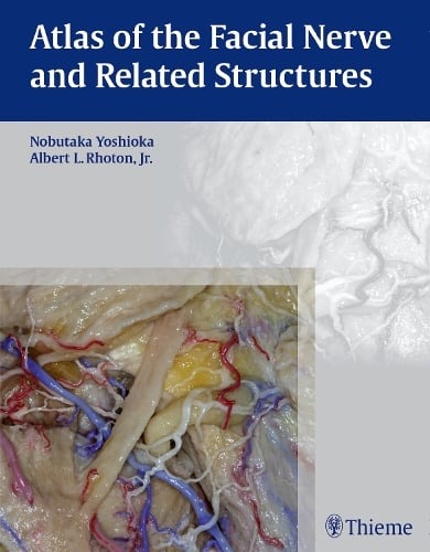
Atlas of the facial nerve and related structures
Opens a unique window into the anatomical intricacies of complex facial nerves and related structures. Special Features: - Exquisite color photographs, prepared from carefully dissected latex injected cadavers, reveal anatomy layer by layer, with remarkable detail and clarity.
Atlas of Regional Anatomy of the Brain Using MRI : with functional correlations
The volume provides a unique review of the essential topographical anatomy of the brain from an MRI perspective, correlating high-quality anatomical plates with the corresponding high-resolution MRI images. The book includes a historical review of brain mapping and an analysis of the essential reference planes used for the study of the human brain. Subsequent chapters provide a detailed review of the sulcal and the gyral anatomy of the human cortex, guiding the reader through an interpretation of the individual brain atlas provided by high-resolution MRI.
Atlas of Neuromuscular Diseases : A Practical Guideline
A comprehensive outline of neuromuscular diseases, written by experienced American and European authors. It discusses all aspects of neuromuscular disorders including the cranial nerves, spinal nerves, motor neurone disease, the nerve plexus, peripheral nerves, mononeuropathies, entrapment syndromes, polyneuropathies, the neuromuscular junction, and muscle disease. Each chapter is uniformly structured into anatomy, symptoms, signs, pathogentic possibilities, diagnosis and differential diagnosis, therapy and prognosis. Additionally the diagnostic tools and investigations used in neuromuscular disease are explained and a practical guide is given how to advance from symptoms to syndromes. For each disease the therapeutic options are described. It contains large number of clinical and histologic pictures from the practical experience of the authors and also a number of artists drawings to facilitate the understanding of anatomic structures.
Atlas of Morphology and Functional Anatomy of the Brain
Divided into a morphological and a functional imaging section. The morphological atlas includes 3D surface images, axial, coronal, and sagittal scans acquired with high-definition T2 fast spin echo (FSE) sequences, and standard and inverted-contrast images. The MR scans are shown side by side with the corresponding anatomical brain sections, provided by Prof. Henri Duvernoy, for more effective comparison. The anatomical nomenclature adopted for both the MR and the anatomical images is listed in an jacket flap for easier consultation.
Atlas of Emergency Medicine Procedures
Provides a step-by-step, visual guide to the most common procedures in emergency medicine. Completely revised, it also includes new procedures such as REBOA, the HINTS test, sphenopalatine ganglion block, occipital nerve block, and lung ultrasonography. Procedures are described on a single page, or two-page spreads, so that the physician can quickly access and review the procedure at hand. The atlas contains more than 700 diagnostic algorithms, schematic diagrams, and photographic illustrations to highlight the breadth and depth of emergency medicine. Topics are logically arranged by anatomic location or by type of procedure, and all procedures are based on the most current and evidence-based practices.
Atlas of electroanatomic mapping in supraventricular arrhythmias
For decades clinical electrophysiology has been considered a discipline for adepts, able to interpret not so obvious intracardiac signals. With the advent of new technologies, offering a three-dimensional reconstruction of cardiac anatomy and generating colour-coded maps, the approach to the complex world of electrophysiology is changing: A three-dimensional colour-coded electroanatomic map of a left atrium could be a precise and self-explanatory representation based on intracavitary signals, on which correct diagnosis is made and successul ablation is performed. As expected, this makes the difference especially in cases exhibiting complex arrhythmias (atypical atrial flutters, postsurgical arrhythmias), in which the task could be really tough.
Atlas of dentomaxillofacial anatomical imaging
A complete guide on imaging of the dentomaxillofacial region, a region of high interest to a wide range of specialists. A large number of injuries and patient’s treatment involve the facial skeleton. Enriched by radiographic images and illustrations, this book explores the anatomy of this region presenting its imaging characteristics through the most commonly available techniques (MDCT, CBCT, MRI and US). In addition, two special chapters on angiography and micro-CT expand the limits of dentomaxillofacial imaging.
Atlas de pathologie thoracique = Atlas of thoracic pathology
This book, devoted primarily to chest imaging, is divided into three main parts. The first brings together all the information to understand the formation and interpretation of images of the thorax, whether normal or pathological, and thus to avoid as many misinterpretations as possible. The second studies the different thoracic radioanatomical syndromes. It allows the pulmonologist, faced with a certain type of image, to relate it to the underlying anatomical compartment damaged and then to consider all the possible etiologies. The third part, on the other hand, recapitulates all the radiological aspects likely to be encountered in each of the pathologies considered.
