



الصفحة 9
الصفحة 9
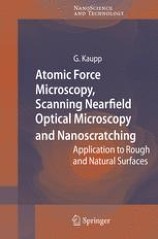
Atomic Force Microscopy, Scanning Nearfield Optical Microscopy and Nanoscratching : Application to Rough and Natural Surfaces
Making a clear distinction is made between nano- and micro-mechanical testing for physical reasons, this monograph describes the basics and applications of the supermicroscopies AFM and SNOM, and of the nanomechanical testing on rough and technical natural surfaces in the submicron range down to a lateral resolution of a few nm. New or improved instrumentation, new physical laws and unforeseen new applications in all branches of natural sciences (around physics, chemistry, mineralogy, materials science, biology and medicine) and nanotechnology are covered as well as the sources for pitfalls and errors.

Atlas of endomicroscopy
Deals with the new insights of endomicroscopy and provides you an overview about the development, the requirements, the technique, current indications and further possibilities of endomicroscopy.

Atlas of confocal laser scanning in-vivo microscopy in ophthalmology : principles and applications in diagnostic and therapeutic
Confocal microscopy with laser scanning technology yields in-vivo images of ocular and ocular adnexal surfaces that are so brilliant that they rival histology in terms of quality.This unique atlas and textbook demonstrates normal in-vivo anatomy of the cornea, limbus and conjunctiva, quantifies various cellular structures using cell-density calculations and establishes correlations between novel optical sections of various diseases of the ocular surface and clinical findings.

Applied scanning probe methodsVII : Biomimetics and industrial applications
The present volumes cover three main areas: novel probes and techniques (Vol. V), charactarization (Vol. VI), and biomimetics and industrial applications (Vol. VII). Volume V includes an overview of probe and sensor technologies including integrated cantilever concepts, electrostatic microscanners, low-noise methods and improved dynamic force microscopy techniques, high-resonance dynamic force - croscopy and the torsional resonance method, modelling of tip cantilever systems, scanning probe methods, approaches for elasticity and adhesion measurements on the nanometer scale as well as optical applications of scanning probe techniques based on near?eld Raman spectroscopy and imaging.

Applied scanning probe methods X : Biomimetics and industrial applications
The success of the Springer Series Applied Scanning Probe Methods I–VII and the rapidly expanding activities in scanning probe development and applications worldwide made it a natural step to collect further speci c results in the elds of development of scanning probe microscopy techniques (Vol. VIII), characterization (Vol. IX), and biomimetics and industrial applications (Vol. X). These three volumes complement the previous set of volumes under the subject topics and give insight into the recent work of leading specialists in their respective elds. Following the tradition of the series, the chapters are arranged around techniques, characterization and biomimetics and industrial applications. Volume VIII focuses on novel scanning probe techniques and the understanding of tip/sample interactions. Topics include near eld imaging, advanced AFM, specialized scanning probe methods in life sciences including new self sensing cantilever systems, combinations of AFM sensors and scanning electron and ion microscopes, calibration methods, frequency modulation AFM for application in liquids, Kelvin probe force microscopy, scanning capacitance microscopy, and the measurement of electrical transport properties at the nanometer scale.
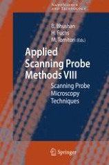
Applied scanning probe methods VIII : Scanning probe microscopy techniques
The success of the Springer Series Applied Scanning Probe Methods I–VII and the rapidly expanding activities in scanning probe development and applications worldwide made it a natural step to collect further speci c results in the elds of development of scanning probe microscopy techniques (Vol. VIII), characterization (Vol. IX), and biomimetics and industrial applications (Vol. X). These three volumes complement the previous set of volumes under the subject topics and give insight into the recent work of leading specialists in their respective elds. Following the tradition of the series, the chapters are arranged around techniques, characterization and biomimetics and industrial applications. Volume VIII focuses on novel scanning probe techniques and the understanding of tip/sample interactions. Topics include near eld imaging, advanced AFM, s- cializedscanningprobemethodsinlifesciencesincludingnewselfsensingcantilever systems, combinations of AFM sensors and scanning electron and ion microscopes, calibration methods, frequency modulation AFM for application in liquids, Kelvin probe force microscopy, scanning capacitance microscopy, and the measurement of electrical transport properties at the nanometer scale.

Applied scanning probe methods VI : Characterization
The scanning probe microscopy feld has been rapidly expanding. It is a demanding task to collect a timely overview of this feld with an emphasis on technical dev- opments and industrial applications. It became evident while editing Vols. I–IV that a large number of technical and applicational aspects are present and rapidly - veloping worldwide. Considering the success of Vols. I–IV and the fact that further colleagues from leading laboratories were ready to contribute their latest achie- ments, we decided to expand the series with articles touching felds not covered in the previous volumes. The response and support of our colleagues were excellent, making it possible to edit another three volumes of the series
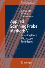
Applied scanning probe methods V : Scanning probe microscopy techniques
The scanning probe microscopy ?eld has been rapidly expanding. It is a demanding task to collect a timely overview of this ?eld with an emphasis on technical dev- opments and industrial applications. It became evident while editing Vols. I–IV that a large number of technical and applicational aspects are present and rapidly - veloping worldwide. Considering the success of Vols. I–IV and the fact that further colleagues from leading laboratories were ready to contribute their latest achie- ments, we decided to expand the series with articles touching ?elds not covered in the previous volumes. The response and support of our colleagues were excellent, making it possible to edit another three volumes of the series.

Applied scanning probe methods IX : Characterization
The success of the Springer Series Applied Scanning Probe Methods I–VII and the rapidly expanding activities in scanning probe development and applications worldwide made it a natural step to collect further speci c results in the elds of development of scanning probe microscopy techniques (Vol. VIII), characterization (Vol. IX), and biomimetics and industrial applications (Vol. X). These three volumes complement the previous set of volumes under the subject topics and give insight into the recent work of leading specialists in their respective elds. Following the tradition of the series, the chapters are arranged around techniques, characterization and biomimetics and industrial applications. Volume VIII focuses on novel scanning probe techniques and the understanding of tip/sample interactions. Topics include near eld imaging, advanced AFM, s- cializedscanningprobemethodsinlifesciencesincludingnewselfsensingcantilever systems, combinations of AFM sensors and scanning electron and ion microscopes, calibration methods, frequency modulation AFM for application in liquids, Kelvin probe force microscopy, scanning capacitance microscopy, and the measurement of electrical transport properties at the nanometer scale.

Applied scanning probe methods IV : Industrial applications
The sc- ning probes emerged as a new - strument for imaging with a p- cision suf?cient to delineate single atoms. At first there were two – the Scanning Tunneling Microscope, or STM, and the Atomic Force Mic- scope, or AFM. The STM relies on electrons tunneling between tip and sample whereas the AFM depends on the force acting on the tip when it was placed near the sample. These were quickly followed by the M- netic Force Microscope, MFM, and the Electrostatic Force Microscope, EFM. The MFM will image a single magnetic bit with features as small as 10nm. With the EFM one can monitor the charge of a single electron.

Applied scanning probe methods III : Characterization
The sc- ning probes emerged as a new - strument for imaging with a p- cision suf?cient to delineate single atoms. At first there were two – the Scanning Tunneling Microscope, or STM, and the Atomic Force Mic- scope, or AFM. The STM relies on electrons tunneling between tip and sample whereas the AFM depends on the force acting on the tip when it was placed near the sample. These were quickly followed by the M- netic Force Microscope, MFM, and the Electrostatic Force Microscope, EFM. The MFM will image a single magnetic bit with features as small as 10nm. With the EFM one can monitor the charge of a single electron.
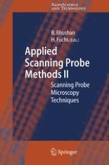
Applied scanning probe methods II : Scanning probe microscopy techniques
The sc- ning probes emerged as a new - strument for imaging with a p- cision suf?cient to delineate single atoms. At first there were two – the Scanning Tunneling Microscope, or STM, and the Atomic Force Mic- scope, or AFM. The STM relies on electrons tunneling between tip and sample whereas the AFM depends on the force acting on the tip when it was placed near the sample. These were quickly followed by the M- netic Force Microscope, MFM, and the Electrostatic Force Microscope, EFM. The MFM will image a single magnetic bit with features as small as 10nm. With the EFM one can monitor the charge of a single electron.

Alzheimers disease : Cellular and molecular aspects of Amyloid beta
This book contains a survey of present-day research into the biomedical fundamentals of Alzheimer’s disease (AD). It contains 20 chapters dealing with widely ranging topics, all of which have a bearing upon the understanding and treatment of AD. Starting with a broad survey of the contribution that the various microscopical techniques (light microscopy, electron microscopy, atomic force microscopy)
Advances in Discrete Tomography and its Applications
Advances in Discrete Tomography and Its Applications is a unified presentation of new methods, algorithms, and select applications that are the foundations of multidimensional image reconstruction by discrete tomographic methods. The self-contained chapters, written by leading mathematicians, engineers, and computer scientists, present cutting-edge research and results in the field.Three main areas are covered: foundations, algorithms, and practical applications. Following an introduction that reports the recent literature of the field, the book explores various mathematical and computational problems of discrete tomography including new applications.

Advanced Time-Correlated Single Photon Counting Techniques
Time-correlated single photon counting (TCSPC) is a remarkable technique for recording low-level light signals with extremely high precision and picosecond-time resolution. TCSPC has developed from an intrinsically time-consuming and one-dimensional technique into a fast, multi-dimensional technique to record light signals. So this reference and text describes how advanced TCSPC techniques work and demonstrates their application to time-resolved laser scanning microscopy, single molecule spectroscopy, photon correlation experiments, and diffuse optical tomography of biological tissue. It gives practical hints about constructing suitable optical systems, choosing and using detectors, detector safety, preamplifiers, and using the control features and optimising the operating conditions of TCSPC devices. Advanced TCSPC Techniques is an indispensable tool for everyone in research and development who is confronted with the task of recording low-intensity light signals in the picosecond and nanosecond range.

Advanced Techniques in Biophysics
Technical advancements are basic elements in our life. In biophysical studies, new applications and improvements in well-established techniques are being implemented every day. This book deals with advancements produced not only from a technical point of view, but also from new approaches that are being taken in the study of biophysical samples, such as nanotechniques or single-cell measurements. This book constitutes a privileged observatory for reviewing novel applications of biophysical techniques that can help the reader enter an area where the technology is progressing quickly and where a comprehensive explanation is not always to be found.

Adsorption and Diffusion
Molecular Sieves - Science and Technology covers, in a comprehensive manner, the science and technology of zeolites and all related microporous and mesoporous materials. Authored by renowned experts, the contributions to this handbook-like series are grouped together topically in such a way that each volume deals with a specific sub-field. Volume 7 is treating fundamentals and analyses of adsorption and diffusion in zeolites including single-file diffusion, i.e. phenomena of basic importance, especially with respect to separation processes and catalysis. Various methods of measuring adsorption and diffusion are described and discussed, i.e. techniques such as chromatographic, gravimetric and barometric uptake and desorption, nuclear magnetic resonance, infrared spectroscopy, interference microscopy, neutron scattering, frequency response as well as proton profiling.

A Guide to Methods in the Biomedical Sciences
A Guide to Methods in the Biomedical Sciences gives a basic description of common methods used in research. This is not intended to be a methods book. Rather, it is intended to be a book that outlines the purpose of the methods described, their limitations and provide alternative approaches as appropriate. Thousands of methods have been developed in the various biomedical disciplines and those covered in this book represent the basic, essential and most widely used methods in several different disciplines. The historical background (including some interesting anecdotes) leading to the development of ground-breaking techniques are described, especially those that significantly advanced the field of biomedical research. Advances that earned their inventors prestigious Nobel Prizes are emphasized. The book is divided into six sections, highlighting selected methods in protein chemistry, nucleic acids, recombinant DNA technology (including forensic based methods), antibody-based techniques, microscopy and imaging, and the use of animals in biomedical sciences.
