الصفحة 9
الصفحة 9
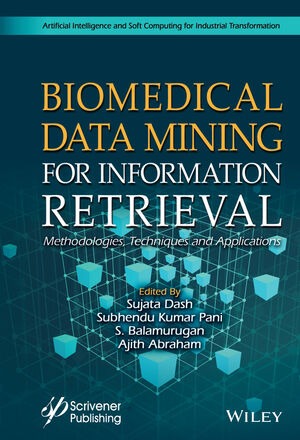
Biomedical data mining for information retrieval : Methodologies, techniques, and applications
Discusses data mining, biomedical image processing, information retrieval with broad coverage of basic scientific applications. covers the topic of mining biomedical text, images and visual features towards information retrieval. Biomedical and health informatics is an emerging field of research at the intersection of information science, computer science, and healthcare and brings tremendous opportunities and challenges due to easily available and abundant biomedical data for further analysis. The aim of healthcare informatics is to ensure the high-quality, efficient healthcare, better treatment and quality of life by analyzing biomedical and healthcare data including patient’s data, electronic health records (EHRs) and lifestyle. Previously, it was a common requirement to have a domain expert to develop a model for biomedical or healthcare; however, recent advancements in representation learning algorithms allows us to automatically to develop the model. Biomedical image mining, a novel research area, due to the vast amount of available biomedical images, increasingly generates and stores digitally.
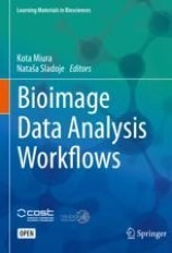
Bioimage Data Analysis Workflows
This book provides students and researchers in the life sciences with essential practical information on how to quantitatively analyze data images. It refrains from focusing on theory, and instead uses practical examples and step-by step protocols to familiarize readers with the most commonly used image processing and analysis platforms such as ImageJ, MatLab and Python.

Big Data in Bioeconomy : Results from the European DataBio Project
This book presents the comprehensive outcome of The European DataBio Project, which examined new data-driven methods to shape a bioeconomy. These methods are used to develop new and sustainable ways to use forest, farm and fishery resources.

Beginning Ubuntu Linux : From novice to professional
Beginning Ubuntu Linux is the perfect guide for those switching to the world's favorite Linux. The new edition has been thoroughly updated to cover technology introduced in the 6.10 release. You'll learn how to install Linux, set up your hardware and software, customize the desktop experience, browse the Web and send/receive e-mail, play back audio and video, edit digital images, use the OpenOffice.org office suite, and more.
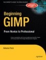
Beginning GIMP : From novice to professional
Beginning GIMP: From Novice to Professional explains how to use the open source image manipulation program, GIMP version 2.4. You'll learn how to install GIMP on Windows, Linux, and MacOS X platforms. Once you've installed the application, you'll learn about the interface and configuration options, and then jump into a quick–and–simple project to familiarize yourself even further. With four–color graphics and screenshots throughout, you'll learn how to prepare camera images for display on web pagesincluding functions like rescaling, cropping, and balancing color. The book also explains with great detail how to utilize layers, paths, and masks. You'll also learn how to draw lines and shapes, use patterns and gradients, and even create your own brushes, patterns, and gradients.

Basic immunology : Functions and disorders of the immune system
Includes recent important advances in our understanding and knowledge of the immune system. A student favorite through six outstanding editions, this new edition uses full-color illustrations and clinical images, useful tables, and practical features such as Summary Point boxes, end-of-chapter review questions, glossary terms, and clinical cases—all designed to help you master this complex topic in the most efficient, effective manner possible.

Basic guide to dental sedation nursing ; 2nd ed.
Starts by defining conscious sedation and discusses why dental sedation is used before moving on to discuss topics such as the medico-legal aspects, the dental nurse’s role, equipment, patient selection, types of sedation, medical emergencies, and anatomy. Presents essential evidence-based information on dental sedation nursing / Reflects the current NEBDN curriculum / Supported by images to demonstrate the concepts discussed

Atlas of Sarcoidosis : Pathogenesis, Diagnosis and Clinical Features
An Atlas of Sarcoidosis: Pathogenesis, Diagnosis and Clinical Features combines illustrations and clinical images of the authors' extensive practices, so that readers have unparalleled access to a comprehensive collection of sarcoidosis images. The atlas is designed to complement and provide a visual supplement to already existing texts on sarcoidosis. Each organ involvement is dealt in a brief and easy to comprehend manner. Various radiographic and laboratory abnormalities are then linked to the clinical features in order to encourage a smooth and easy practical integration at the bedside and to help practising pulmonologists, dermatologists and other clinicians who require a comprehensive visual encyclopedia of sarcoidosis images.

Atlas of Regional Anatomy of the Brain Using MRI : with functional correlations
The volume provides a unique review of the essential topographical anatomy of the brain from an MRI perspective, correlating high-quality anatomical plates with the corresponding high-resolution MRI images. The book includes a historical review of brain mapping and an analysis of the essential reference planes used for the study of the human brain. Subsequent chapters provide a detailed review of the sulcal and the gyral anatomy of the human cortex, guiding the reader through an interpretation of the individual brain atlas provided by high-resolution MRI.

Atlas of Psoriatic Arthritis
A visual guide to the complex and inter-related diseases of psoriatic arthritis and psoriasis, this volume contains more than 150 images spanning a wide spectrum of variations of the disease.

Atlas of Operative Oral and Maxillofacial Surgery
Comprised of concise text and detailed vignettes focusing on surgical indications, contraindications, pertinent anatomy, virtual surgical planning, operative techniques, postoperative management, complications and key points with over 2,000 high-quality images. Serves as an innovative, multidisciplinary, surgical atlas covering core aspects of oral and maxillofacial surgery, head and neck reconstructive surgery, and facial cosmetic surgery. Chapters are written by experts in their fields and are designed to provide high-yield information utilizing a case report format.

Atlas of Morphology and Functional Anatomy of the Brain
Divided into a morphological and a functional imaging section. The morphological atlas includes 3D surface images, axial, coronal, and sagittal scans acquired with high-definition T2 fast spin echo (FSE) sequences, and standard and inverted-contrast images. The MR scans are shown side by side with the corresponding anatomical brain sections, provided by Prof. Henri Duvernoy, for more effective comparison. The anatomical nomenclature adopted for both the MR and the anatomical images is listed in an jacket flap for easier consultation.

Atlas of fallen dust in Kuwait
Serves as an atlas of deposited dust and dust storms in Kuwait in relation to local and global regions. It features a wealth of maps and images of dust storm trajectories in the region, together with detailed descriptions of the chemical and physical properties of fallen dust

Atlas of dentomaxillofacial anatomical imaging
A complete guide on imaging of the dentomaxillofacial region, a region of high interest to a wide range of specialists. A large number of injuries and patient’s treatment involve the facial skeleton. Enriched by radiographic images and illustrations, this book explores the anatomy of this region presenting its imaging characteristics through the most commonly available techniques (MDCT, CBCT, MRI and US). In addition, two special chapters on angiography and micro-CT expand the limits of dentomaxillofacial imaging.

Atlas of confocal laser scanning in-vivo microscopy in ophthalmology : principles and applications in diagnostic and therapeutic
Confocal microscopy with laser scanning technology yields in-vivo images of ocular and ocular adnexal surfaces that are so brilliant that they rival histology in terms of quality.This unique atlas and textbook demonstrates normal in-vivo anatomy of the cornea, limbus and conjunctiva, quantifies various cellular structures using cell-density calculations and establishes correlations between novel optical sections of various diseases of the ocular surface and clinical findings.

Atlas de pathologie thoracique = Atlas of thoracic pathology
This book, devoted primarily to chest imaging, is divided into three main parts. The first brings together all the information to understand the formation and interpretation of images of the thorax, whether normal or pathological, and thus to avoid as many misinterpretations as possible. The second studies the different thoracic radioanatomical syndromes. It allows the pulmonologist, faced with a certain type of image, to relate it to the underlying anatomical compartment damaged and then to consider all the possible etiologies. The third part, on the other hand, recapitulates all the radiological aspects likely to be encountered in each of the pathologies considered.
Astronomy with a home computer
Here is a one-volume guide to just about everything computer-related for amateur astronomers!Today’s amateur astronomy is inextricably linked to personal computers. Computer-controlled "go-to" telescopes are inexpensive. CCD and webcam imaging make intensive use of the technology for capturing and processing images. Planetarium software provides information and an easy interface for telescopes. The Internet offers links to other astronomers, information, and software. The list goes on and on.
Artificial intelligence techniques for satellite image analysis
The main objective of this book is to provide a common platform for diverse concepts in satellite image processing. In particular it presents the state-of-the-art in Artificial Intelligence (AI) methodologies and shares findings that can be translated into real-time applications to benefit humankind. Interdisciplinary in its scope, the book will be of interest to both newcomers and experienced scientists working in the fields of satellite image processing, geo-engineering, remote sensing and Artificial Intelligence. It can be also used as a supplementary textbook for graduate students in various engineering branches related to image processing.
Artificial intelligence in recognition and classification of astrophysical and medical images
This book presents innovative techniques in Recognition and Classification of Astrophysical and Medical Images. The contents include: Introduction to pattern recognition and classification in astrophysical and medical images. Image standardization and enhancement. Region-based methods for pattern recognition in medical and astrophysical images. Advanced information processing using statistical methods. Feature recognition and classification using spectral method
Artificial intelligence for multisource geospatial information
Collects 10 original research contributions published in the Special Issue entitled “Artificial Intelligence for Multisource Geospatial Information” of the ISPRS International Journal of Geo-Information. The focus is on different methods of Geospatial Artificial Intelligence (GeoAI) based on deep learning using different network architectures, clustering, soft computing, and semantic approaches. They are proposed to deal with a variety of Geospatial Big Data (GBD), such as georeferenced texts and photos in social networks, remote sensing images, cartographic maps, multidimensional geo databases, metadata in spatial data infrastructures, and for different tasks, such as for multisource georeferenced text integration and geodata flexible querying, for social sensing by applying sentiment analysis, clustering and geo analysis, for segmentation of roads, clouds and snow, and for detection of small targets and people on the streets.
