الصفحة 2
الصفحة 2

Essentials of Restenosis : For the Interventional Cardiologist
Essentials of Restenosis provides the practicing cardiologists and biologists an elaborate review of the current advances in the field. The book explore the process of restenosis from bench to bedside, encompassing in equal detail the newest scientific findings of the biological mechanisms as well as the progression in development of diagnostics and treatments. The volume also provides the reader with a basic understanding of the biological process and the latest diagnostics and treatments of the disease, with an up-to-date detailed discussion of the most important recent findings.

Dynamic Radiology of the Abdomen : Normal and Pathologic Anatomy
Meyers' Dynamic Radiology of the Abdomen, extensively revised and updated, is the classic text covering radiology of the abdomen as it relates to the progression of disease within an organ and from one organ to another. The book provides a systematic application of anatomic and dynamic principles to the practical understanding and diagnosis of intraabdominal disease. The full range of imaging modalities is addressed, from plain films and conventional contrast studies to CT, US, MRI and endoscopic ultrasonography. Highly selected, ample images including CT and MRI support the thoroughly descriptive text. Expanded references, citing both the classic and recent contributions, and a detailed cross-referenced index are presented. For radiologists, general surgeons, gastroenterologists, and others seeking insight into the clinical practice of radiology, this text continues to be the gold standard in the field.

Dynamic Contrast-Enhanced Magnetic Resonance Imaging in Oncology
Dynamic contrast-enhanced MRI is now established as the methodology of choice for the assessment of tumor microcirculation in vivo. This is assisting clinical practitioners in the management of patients with solid tumors and is finding prominence in the assessment of tumor treatments, including anti-angiogenics, chemotherapy, and radiotherapy. In this book, targeted at both clinical practitioners and basic scientists, the principles of the methods, their practical implementation, and their application to specific tumor types are discussed by the leading authorities in the field today. The book will serve as an invaluable single-volume reference covering all the latest developments in contrast-enhanced oncological MRI.

Diseases of the Heart, Chest & Breast : Diagnostic Imaging and Interventional Techniques
This book deals with imaging of diseases of heart, chest and breast. These fields have substantially advanced during the last few years, driven by both clinical developments and advances in imaging technology.لإheir chapters are disease-oriented and cover all the relevant imaging modalities, including standard radiography, CT, nuclear medicine with PET, ultrasound and magnetic resonance imaging, as well as imaging-guided interventions. As a result, this book presents a comprehensive review of current knowledge in imaging of the heart and chest , as well as thoracic interventions and a selection of "hot topics" of breast imaging.

Color Doppler US of the Penis
This book provides a comprehensive reference and practical guide on the application of US to penile diseases and conditions. The topics covered include erectile dysfunction, Peyronie’s disease, priapism, trauma, tumors, the postoperative penis, inflammation, and fibrosis.
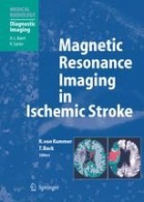
Magnetic Resonance Imaging in Ischemic Stroke
The imaging of stroke has undergone significant changes owing to the rapid progress in imaging technology. This volume, comprising three parts, is designed to provide a comprehensive summary of the current role of MR imaging in patients with ischemic stroke. The first part outlines the clinical presentations of stroke and discusses the diagnostic efficacy and therapeutic impact of MR imaging. The second and third parts form the core of the volume, and are based on a novel approach in that the topic is presented from two very different viewpoints. Part 2 provides a detailed presentation of the distinguishing features of stroke from the radiologist's perspective. By contrast, part 3 addresses the needs of the clinician, documenting specific stroke syndromes and their correlates on MR imaging. The overall aim has been to create a well-illustrated volume with broad appeal that links pathology, radiology and stroke medicine in an informative manner.
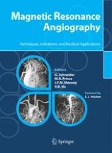
Magnetic resonance angiography : techniques, indications and practical applications
The advent of contrast-enhanced MRA in the early to mid 1990s revolutionized the clinical approach to vascular imaging: an accurate non-invasive imaging modality, not requiring ionizing radiation or potentially nephrotoxic iodinated contrast media, was able to compete with the more hazardous and invasive catheter angiography. Today, MRA is a safe, easy-to-perform procedure routinely used in most imaging centers, and the continued development of faster, more powerful magnets and more effective contrast agents is increasingly helping to overcome many of the early limitations of the technique.
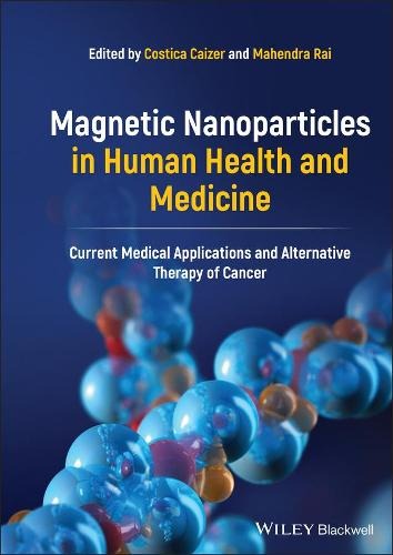
Magnetic nanoparticles in human health and medicine : Current medical applications and alternative therapy of cancer
Progress in improving diagnosis by magnetic resonance imaging (MRI) and using non-invasive and non-toxic magnetic nanoparticles for targeted drug delivery. Focusing on cancer diagnosis and therapy, the book covers both fundamental principles and advanced theoretical and experimental research on the magnetic properties, biocompatibilization, biofunctionalization, and application of magnetic nanoparticles in nanobiotechnology and nanomedicine.
Machine learning for neurodegenerative disorders : advancements and applications
Explores the application of machine learning to the understanding, early diagnosis, and management of neurodegenerative disorders. With a specific focus on its role in ongoing clinical trials, the book covers essential topics such as data collection, pre-processing, feature extraction, model development, and validation techniques. It delves into the applications of neuroimaging techniques like magnetic resonance imaging (MRI), computed tomography (CT), and positron emission tomography (PET) in the diagnosis and understanding of neurodegenerative disorders. Additionally, the book examines various machine-learning algorithms employed for biomarker discovery in neurodegenerative disorders. It highlights the role of neuroinformatics and big data analysis in advancing the understanding and management of neurodegenerative disorders. Furthermore, the book reviews future prospects and presents the ethical considerations and regulatory challenges associated with implementing machine learning approaches in the diagnosis, treatment, and prevention of neurodegenerative disorders.

Clinical MR Imaging : A Practical Approach
This book offers practical guidelines for performing efficient and cost-effective MRI examinations in daily practice. The underlying idea is that, by adopting a practical protocol-based approach, the work-flow in a MRI unit can be streamlined and optimized.
Classic Papers in Modern Diagnostic Radiology
The subject of diagnostic radiology is now very large and radiology depa- ments are involved in all areas of modern patient care.The defining event in m- ern radiology,and arguably the most significant development in radiology since Wilhelm Röntgen discovered X-rays, was the invention of the CT scanner in the 1970s.The CT scanner introduced modern cross-sectional imaging and also di- tal imaging.We now have MRI and ultrasound and these techniques are replacing many traditional X-ray procedures.The developments in radiology have been the result of a fruitful interaction between the basic sciences, clinical medicine and the manufacturers. This can be seen by looking at the various sources of these publications. Change is produced by the interactions between the various dis- plines. The editors have had a very difficult task in selecting the key discoveries and descriptions.The radiological literature is very large.Medical imaging continues to develop rapidly and these papers are the foundations of our current practice.
Cardiovascular MRI in practice : A teaching file approach
This text resource outlines the systematic approach to CMR interpretation. The depiction of a "core exam" and the modifications used for a variety of patient circumstances are demonstrated using simple visual assessment of the images. Special emphasis on the advantages of CMR relative to other modalities reinforces practical learning objectives, organized so that the reader starts with patient images – as one would in a clinical scenario – and works back to the didactic material.
Breast cancer ; 2nd ed.
This book highlights M. D. Anderson Cancer Center’s multidisciplinary approach and reviews the entire spectrum of patient care, from prevention and screening through diagnosis and treatment through posttreatment follow-up and survivorship issues.
Bone densitometry for technologists
Radiologic technologists, nurse practitioners, physician assistants, and dedicated densitometry technologists can find new guidelines for bone density testing, new therapies for osteoporosis, and new treatment guidelines for osteoporosis, as well as new chapters on pediatric densitometry body composition assessments, and the use of skeletal morphometry in diagnosis and fracture risk prediction. The twelve appendices have also been updated to reflect the most current information available, including contacts for densitometry equipment manufacturers and organizations, guidelines for bone density testing and CPT codes, definitions of terms, and new conversion equations.

Atlas of topographical and pathotopographical anatomy of the head and neck
Presents the ultrasonic topographical and pathotopographical anatomy of the head and neck, offering further detail into these important areas for use by medical professionals. This atlas of topographic and pathotopographic human anatomy is a fundamental and practically important book designed for doctors of all specializations and students of medical schools. Here you can find almost everything that is connected with the topographic and pathotopographic human anatomy, including original graphs of logical structures of topographic anatomy and development of congenital abnormalities, topography of different areas in layers, pathotopography, computer and magnetic resonance imaging (MRI) of topographic and pathotopographic anatomy.

Atlas of Morphology and Functional Anatomy of the Brain
Divided into a morphological and a functional imaging section. The morphological atlas includes 3D surface images, axial, coronal, and sagittal scans acquired with high-definition T2 fast spin echo (FSE) sequences, and standard and inverted-contrast images. The MR scans are shown side by side with the corresponding anatomical brain sections, provided by Prof. Henri Duvernoy, for more effective comparison. The anatomical nomenclature adopted for both the MR and the anatomical images is listed in an jacket flap for easier consultation.
Alternative breast imaging : Four model-based approaches
Medical imaging has been transformed over the past 30 years by the advent of computerized tomography (CT), magnetic resonance imaging (MRI), and various advances in x-ray and ultrasonic techniques. An enabling force behind this progress has been the (so far) exponentially increasing power of computers, which has made it practical to explore fundamentally new approaches. In particular, what our group terms "model-based" modalities-which produce tissue property images from data using nonlinear, iterative numerical modeling techniques-have become increasingly feasible. Alternative Breast Imaging: Four Model-Based Approaches explores our research on four such modalities, particularly with regard to imaging of the breast: (1) MR elastography (MRE), (2) electrical impedance spectroscopy (EIS), (3) microwave imaging spectroscopy (MIS), and (4) near infrared spectroscopic imaging (NIS).

Advances in Medical Engineering
In this book, research and development trends of physics, engineering, mathematics and computer sciences in biomedical engineering are presented. Contributions from industry, clinics, universities and research labs with foci on medical imaging (CT, MRT, US, PET, SPECT etc.), medical image processing (segmentation, registration, visualization etc.), computer-assisted surgery (medical robotics, navigation), biomechanics (motion analysis, accident research, computer in sports, ergonomics etc.), biomedical optics (OCT, soft-tissue optics, optical monitoring etc.) and laser medicine (tissue ablation, gas analytics, topometry etc.) give insight to recent engineering, clinical and mathematical studies.
Advanced Bioimaging Technologies in Assessment of the Quality of Bone and Scaffold Materials : Techniques and Applications
This book provides a perspective on the current status of bioimaging technologies developed to assess the quality of musculoskeletal tissue with an emphasis on bone and cartilage. It offers evaluations of scaffold biomaterials developed for enhancing the repair of musculoskeletal tissues. These bioimaging techniques include micro-CT, nano-CT, pQCT/QCT, MRI, and ultrasound, which provide not only 2-D and 3-D images of the related organs or tissues, but also quantifications of the relevant parameters. The advance bioimaging technologies developed for the above applications are also extended by incorporating imaging contrast-enhancement materials.
Adrenal Glands : Diagnostic Aspects and Surgical Therapy
Surgery of the Adrenal Glands is a comprehensive medical textbook that includes everything that a surgeon, but for all purposes any physicain or medical student, may want to find on adrenal surgery in a friendly, up to date, evidence based manner. 30 of the most renowned names in the field of adrenal surgery from the US and Europe draw on their experience in exploring diagnosis, indications for surgery, choice of operation and operative techniques, and surgical outcomes for every major adrenal condition. Over 200 exquisite illustrations, ranging from intraoperative photographs, line drawings, diagnostic images, pathology slides, tables and graphs are available to make each case discussed comprehensible and easy to follow. Traditional and novel therapeutic recommendations are introduced and the latest developments in minimal-access operative techniques are discussed and reviewed. Finally, the best approaches in dealing with challenging conditions such as adrenal incidentaloma, subclinical Cushing's syndrom, Congenital Adrenal Hyperplasia, Mineralcorticoid Express Syndrome, Extraadrenal and Malignant Pheochromocytoma are discussed.
