


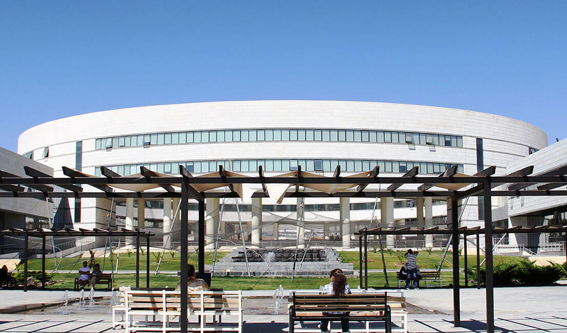
الصفحة 1
الصفحة 1

Maxillofacial Imaging
Demonstrates how advanced medical imaging techniques can be successfully applied to dental and maxillofacial conditions. There is a focus on CT and MRI, but the use of all contemporary imaging techniques are illustrated including PET, PET/CT, ultrasonography, and cone beam CT. The presentation is in atlas style, with succinct, bulleted text and a wealth of high-quality images in multiple planes. All images for each patient are grouped to enable the reader very quickly to gain an imaging overview of the condition under consideration. After a comprehensive introductory chapter on normal imaging anatomy, the role of advanced imaging techniques is described in pathologic conditions of the mandible and maxilla, temporomandibular joint, regions closely related to the jaw, paranasal sinuses, oral cavity, salivary glands, and structures adjacent to the maxillofacial region. A concluding chapter examines the use of interventional procedures for diagnosis and treatment of maxillofacial conditions. Compared with the first edition, numerous additional cases have been incorporated and a completely new chapter focuses on cone beam CT.

Implantology step by step
A consistently practice-oriented, instruction- based and illustrated technical textbook, has provided guidance and help to a whole generation of dentists venturing into the field of dental implant treatment. Brought together the treatment of the maxilla and mandible in one volume and has included a chapter on the totally edentulous patient. A wealth of newly included patient reports illustrate novel methods and techniques, replacing any material that has since become obsolete. What remains is the innovative concept that a variety of baseline situations and degrees of difficulty are taken into account for all clinical situations - from the restoration of singletooth gaps to the treatment of the edentulous maxilla and mandible with sinus elevation and immediate implants. Important complications are also considered, with causes and solutions.

Illustrated Pediatric Dentistry ; Part 1
This textbook is modernized with the latest information and techniques in pediatric dentistry. The chapters cover primary pediatric dentistry, its clinical aspects, preventive dentistry, and information about the latest trends in the specialty. The text will equip readers with the knowledge suited to the changing
Clinical oral anatomy : A comprehensive review for dental practitioners and researchers
Presents the most current and comprehensive review of oral anatomy for clinicians and researchers alike. In 26 chapters, the reader is taken on a unique anatomical journey, starting with the oral fissure, continuing via the maxilla and mandible to the tongue and floor of the mouth, and concluding with the temporomandibular joint and masticatory muscles. Each chapter offers a detailed description of the relevant anatomical structures and their spatial relationships, provides quantitative morphological assessments, and explains the relevance of the region for clinical dentistry. All dental health care professionals require a sound knowledge of anatomy for the purposes of diagnostics, treatment planning, and therapeutic intervention. A full understanding of the relationship between anatomy and clinical practice is the ultimate objective, and this book will enable the reader to achieve such understanding as the basis for provision of the best possible treatment for each individual patient as well as recognition and comprehension of unexpected clinical findings.
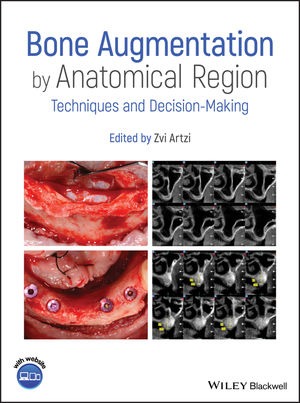
Bone augmentation by anatomical region : Techniques and decision-making
It's a unique, evidence-based guide focusing on each specific anatomical region – anterior maxilla, posterior maxilla, anterior mandible, and posterior mandible – in order to emphasize the correct implemented procedures needed to successfully perform oral osseous reconstruction.

Atlas of mandibular and maxillary reconstruction with the fibula flap : A step-by-step approach
Deals with the standard technique used for reconstructing the mandible and the maxilla - the fibula flap. The reader will find useful information on all issues that are important in the surgical procedure, including the use of CAD-CAM technology (Computer Assisted Technology), bone synthesis and flap modelling. The editors draw on their 30 years of experience to provide a step-by-step description of this surgical procedure. With the help of numerous illustrations, the reader will learn the technical, functional and aesthetic developments since 1989 when this technique was first described.

Assessment of mandibular angle and condyle morphology in a sample of Syrian people
The purpose of this study was to determine the prevalence of radiographic changes in the mandibu‐ lar angle (bone apposition) and osseous alterations in the temporomandibular joints (TMJs) in a sample of adult Syrian population . In addition, the study intended to investigate possible correlations between the two sites of contour bone changes (mandibular angle and TMJ) and to analyze various patient‐related factors, including sex, age and dental status.
Artificial intelligence applied to medical imaging and computational biology
Medical imaging and computational biology continuously pose new fundamental medical and biological questions that often give rise to novel challenges in Artificial Intelligence. These research fields present an increasing need for the application of cutting-edge computational approaches that generally involve machine learning or computational intelligence techniques, which can effectively perform bioimage and biosignal processing in different clinical areas.
