
الصفحة 6
الصفحة 6

Imaging of the liver and Intra-hepatic biliary tract ; Vol.2 : Tumoral pathologies
This is the second of two volumes that together provide a comprehensive analysis of the embryology, normal anatomy, and pathology of the liver and intrahepatic biliary tract as seen on modern diagnostic imaging techniques. In this second volume, readers will find comprehensive description and illustration of the imaging appearances of tumoral pathologies, both in the “normal liver” and in the context of chronic liver disease and liver cirrhosis. In addition, the imaging findings in relation to different treatment approaches are presented, with extensive coverage of imaging of tumor response and post-treatment changes.

Imaging of the liver and Intra-hepatic biliary tract ; Vol.1 : Imaging techniques and non-tumoral pathologies
This is the first of two volumes that together provide a comprehensive analysis of the embryology, normal anatomy, and pathology of the liver and intrahepatic biliary tract as seen on modern diagnostic imaging techniques. In this volume, readers will find fundamental information on embryology, radiological anatomy, and anatomic variants. A thorough introduction is then provided to each imaging technique, including ultrasound, computed tomography, magnetic resonance imaging, nuclear medicine techniques, angiography, and interventional radiology. The remainder of the volume is devoted to non-tumoral pathology of the liver and intra-hepatic biliary tract

Imaging of Parasitic Diseases
This book provides an overview of the imaging findings of parasitic diseases using modern imaging equipment. The chapters consist of short descriptions of causative pathogens, epidemiology, modes of transmission, pathology, clinical manifestations, laboratory tests, and imaging findings.

Imaging of Orthopedic Sports Injuries ; 2nd ed.
This book, now in a revised and updated second edition, provides a review of imaging abnormalities in orthopedic sports injuries. The first part of the book includes background information on relevant basic science and general imaging principles in sports traumatology, while the second part discusses the topography of various sports injuries. Each chapter highlights the merits of different imaging techniques, focusing on a specific clinical problem. The third part then examines natural history and monitoring. Several new chapters have been added, including a chapter on postoperative joint imaging in the sports patient as well as image-guided interventions in sports injuries.

Imaging of Orthopedic Sports Injuries ; 1st ed.
This volume provides an updated review of imaging abnormalities in orthopedic sports injuries. The first part of the book contains background information on relevant basic science and general imaging principles in sports traumatology. The second part comprises a topographic discussion of sports injuries. Each chapter highlights the merit of different imaging techniques, focused on a specific clinical problem. In the third part, natural history, monitoring and follow-up by imaging are discussed. This well-illustrated book will be of value for musculoskeletal radiologists, orthopedic surgeons, sports physicians and everyone else involved in sports medicine.

Imaging of Orbital and Visual Pathway Pathology
This is one of the first books to deal with imaging of pathology of the entire visual system. It is divided into two parts, general and special. In the general part, the most important basics of modern imaging methods are discussed in detail, but with less emphasis on the physical background than in purely neuro-/radiological books. A chapter is devoted to the meticulous presentation of imaging anatomy of the orbit and intracranial visual pathway.

Imaging of occupational and environmental disorders of the chest
This book provides an up-to-date and comprehensive approach to modern imaging of environmental and occupational diseases of the chest. The first part of the book addresses the basic knowledge required to understand imaging in this context, while the second focuses on the imaging results achieved in a variety of specific disorders. There is particular emphasis on the role of thin-section computed tomography since this technique facilitates the detection of early subclinical abnormalities.

Imaging of Kidney Cancer
This is one of the first books to deal specifically with diagnostic imaging of the entire spectrum of kidney cancers. Both new and conventional imaging modalities are fully considered. In an introductory chapter, the histopathological classification of kidney cancers is presented and discussed. The following five chapters describe in great detail the abilities, advantages, and disadvantages of the various imaging modalities used in the diagnosis of kidney cancers and the assessment of disease extension. Subsequent chapters offer an exhaustive description of the radiological features of the different histological subtypes of kidney cancer, with radiological and histological illustrations and tables.

Imaging nelle urgenze vascolari - Body : Casi clinici = Imaging in vascular emergencies - Body : Clinical cases
Vascular urgency is true urgency. Given the variability of clinical presentations, the radiologist is increasingly called into question for diagnostic-differential problems and he is entrusted with the nosological framework and the therapeutic choices of doubtful cases. The purpose of the book is to provide, through the presentation of a large number of cases, an easy, fast and stimulating means through which the reader can confront.

Imaging in Treatment Planning for Sinonasal Diseases
Provides both basic and advanced information on the clinical presentation, imaging findings and treatment of sinonasal diseases. For each specific disease, the rationale underlying the treatment strategy is discussed and the imaging findings critical to the decision-making process are identified. This is an original approach and reflects the constant team effort at the editors' institute over the past 15 years to integrate clinical and radiological information with the aim of establishing the most appropriate treatment. Special emphasis is placed on the identification of clinical and imaging data that allow selection of an endonasal or an external approach in surgical candidates. The value of planning an integrated endoscopic and radiological follow-up in the different diseases is also thoroughly reviewed. Through the correlation of imaging with endoscopic or clinical findings, this book will enable radiologists to familiarise themselves with the "otorhinolaryngological world" and clinicians to understand more fully the significance of specific radiological findings. It will serve as an invaluable guide to the selection of imaging techniques.

Imaging in Transplantation
This book covers all topics related to the imaging of organ transplantation. An introductory section addresses such issues as organ procurement, patient selection, immune responses, and ethical and economic considerations. The main part of the book then offers in-depth coverage of heart, renal, liver, lung, bone marrow and pancreatic and intestinal transplantation.

Imaging in Pediatric Skeletal Trauma : Techniques and Applications
This is a comprehensive textbook on imaging of pediatric skeletal trauma. It provides a detailed description of the techniques used and the imaging findings, detailing their clinical relevance. Emphasis is placed on those injuries and their radiological features which are important to the orthopedic surgeon and the successful management of the child.

Imaging in Pediatric Dental Practice : A Guide to Equipment, Techniques and Clinical Considerations
This book is a comprehensive guide to dentomaxillofacial imaging in paediatric dentistry and is an excellent resource for both general dental practitioners and paediatric dentists.

Imaging in Oncology
Imaging in Oncology consists of scholarly reviews that describe the role of imaging in oncology for diagnosis, follow-up and image guided interventions. Experts in various fields of radiology have contributed to this book.
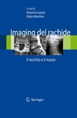
Imaging del rachide : Il vecchio e il nuovo = Spine Imaging: The Old and the New
The vertebral column represents the anatomical district most subjected to diagnostic imaging investigations and, in this field, much of the "old" still retains its relevance. The volume fits into this context as a valid professional refresher tool aimed at the radiologist, but is also a useful reference work for the orthopedist, rheumatologist and physiatrist.

Imaging Atlas of the Pelvic Floor and Anorectal Diseases
The goal of Imaging Atlas of the Pelvic Floor and Anorectal Diseases is to clearly and precisely present indications, techniques, limitations, sources of errors, and pitfalls of these imaging modalities.

Image-Guided Interventions : Technology and Applications
Responding to the growing demand for minimally invasive procedures, Image-Guided Interventions: Technology and Applications provides a cohesive overview of the current technological advances in image-guided surgery, and their applications in the clinical environment.

Image Processing in Radiology : Current Applications
Few fields have witnessed such impressive advances as image processing in radiology. The progress achieved has revolutionized diagnosis and greatly facilitated treatment selection and accurate planning of procedures. This book provides a comprehensive and up-to-date description of how to use 2D and 3D processing tools in clinical radiology.

Ileo meccanico dell’intestino tenue : Aspetti TC e correlazioni eco-radiografiche = Mechanical ileus of the small intestine : CT aspects and echo-radiographic correlations
Mechanical occlusion of the small intestine is a frequently encountered clinical problem, whose clinical-instrumental management is made difficult by the complexity of the mechanisms responsible for the occlusive picture. The role of the radiologist is to promptly guide the therapeutic process on the basis of a correct interpretation of the iconographic pictures. The purpose of this volume - the only one of its kind - is to relate the pathophysiology of occlusion to imaging. The rich iconography, which ranges from clinical onset to resolution, emphasizes the role of multidetector computed tomography with intravenous contrast, which is the current gold standard. However, the correlations with the echo-radiographic methods are not forgotten, which retain an effective diagnostic role, with a low biological and economic cost. The mechanical ileus of the small intestine is a striking example in support of the dream of many professionals: that, that is, to have a figure of a fine abdominal emergency radiologist, an expert in organ and disease and not only in the machine, as often unfortunately it happens.

IAEA Atlas of Cardiac PET/CT : A Case-Study Approach
This book presents a wide portfolio of examples of positron emission tomography coupled with computer tomography (PET/CT) studies in various cardiac conditions in order to provide a rationale for the implementation of this technology in an array of clinical conditions. Cardiovascular diseases are a major contributor to premature morbidity and mortality worldwide. Low- and middle-income countries (LMICs) are particularly affected by cardiovascular diseases (CVDs), with more than 75% of all CVDs deaths occurring in these countries. For this reason, target 3.4 of the United Nations (UN) Sustainable Development Goals (SDGs) agenda aims at a 30% reduction in premature mortality due to non-communicable diseases (NCDs), which include CVDs, by 2030.
